. 2019 Jan 7;19(1):19.
doi: 10.1186/s12885-018-5214-8.

Differences in tumor microenvironments between primary lung tumors and brain metastases in lung cancer patients: therapeutic implications for immune checkpoint inhibitors

Affiliations

Differences in tumor microenvironments between primary lung tumors and brain metastases in lung cancer patients: therapeutic implications for immune checkpoint inhibitors

Ryul Kim et al. BMC Cancer. .

Abstract

Background: We aimed to compare intra- and extracranial responses to immune checkpoint inhibitors (ICIs) in lung cancer with brain metastases (BM), and to explore tumor microenvironments of the brain and lungs focusing on the programmed cell death-1 (PD-1)/programmed cell death ligand-1 (PD-L1) pathway.

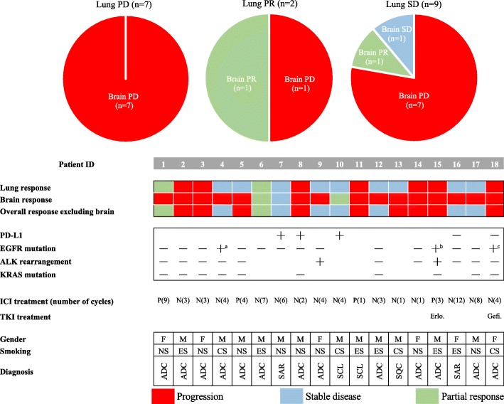
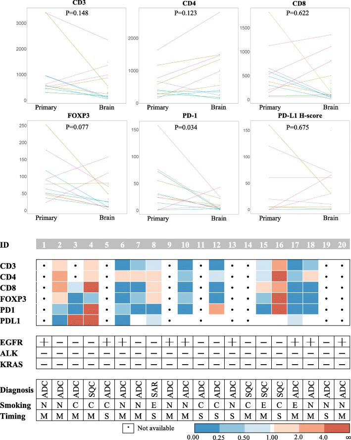
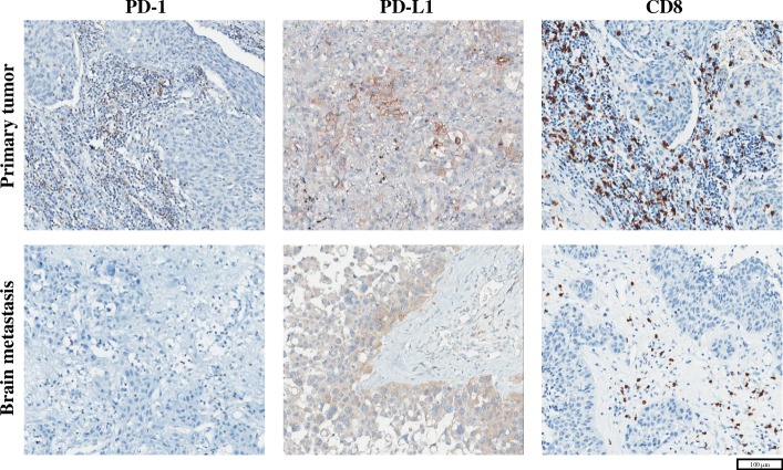
Methods: Two cohorts of lung cancer patients with BM were analyzed. Cohort 1 included 18 patients treated with nivolumab or pembrolizumab, and intra- and extracranial responses were assessed. Cohort 2 comprised 20 patients who underwent both primary lung surgery and brain metastasectomy. Specimens from cohort 2 were subjected to immunohistochemical analysis for the following markers: CD3, CD4, CD8, FOXP3, and PD-1 on tumor infiltrating lymphocytes (TIL) and PD-L1 on tumor cells.

Results: Seven patients (38.9%) in cohort 1 showed progressive disease in both primary and intracranial lesions. Although the other 11 patients exhibited a partial response or stable disease in the primary lesion, eight showed a progression in BM. Interestingly, PD-1+ TILs were significantly decreased in BM (P = 0.034). For fifteen patients with adenocarcinoma, more distinctive patterns were observed in CD3+ (P = 0.078), CD8+ (P = 0.055), FOXP3+ (P = 0.016), and PD-1+ (P = 0.016) TILs.

Conclusions: There may be discordant responses to an ICI of lung cancer between primary lung lesion and BM based on discrepancies in the tumor microenvironment. The diminished infiltration of PD-1+ TILs in tumor tissue within the brain may be one of the major factors that hinder the response to anti-PD-1 antibody in BM.

Keywords: Brain metastasis; Immunotherapy; Lung cancer; PD-1; PD-L1.


Conflict of interest statement

Ethics approval and consent to participate

This study was approved by the SNUH Institutional Review Board (IRB approval number: H-1702-158-836) and was conducted in accordance with Declaration of Helsinki provisions. Written informed consent to participate in the study was obtained from each patient at admission.

Consent for publication

Not applicable.

Competing interests

The authors declare that they have no competing interests.

Publisher’s Note

Springer Nature remains neutral with regard to jurisdictional claims in published maps and institutional affiliations.

Figures

Fig. 1
Fig. 1
Treatment response to anti–PD-1 antibody in cohort 1. (Upper) Pie charts demonstrating the response in primary lung cancer and paired brain metastases. (Lower) The treatment response along with clinicodemographic characteristics of individual patients in cohort 1. Each patient received either nivolumab (N) or pembrolizumab (P) for the specified number of cycles. Each tile in this figure denotes the treatment response: red for progressive disease, blue for stable disease, and green for a partial response. Abbreviations: PD, progressive disease; PR, partial response; SD, stable disease; ID, identification number; P, pembrolizumab; N, nivolumab; PD-L1, programmed cell death ligand-1; PD-1, programmed cell death-1; M, male; F, female; EGFR, epidermal growth factor receptor; ALK, anaplastic lymphoma kinase; NS, never smoked; ES, ex-smoker; CS, current smoker; ADC, adenocarcinoma; SAR, pulmonary sarcomatoid carcinoma; SCL, small-cell lung cancer; SQC, squamous cell carcinoma, Erlo., erlotinib; Gefi., gefitinib; ICI, immune checkpoint inhibitor; TKI, tyrosine kinase inhibitor. a: Exon 20 insertion. b: Exon 19 deletion. c: L858R mutation
Fig. 2
Fig. 2
Immunohistochemical analysis of CD3, CD4, CD8, FOXP3, and PD-1 on tumor-infiltrating lymphocytes, and PD-L1 on tumor cells of patients in cohort 2. (Upper) Ladder plots demonstrate the different expression of each marker between primary lung cancer and brain metastases. Individual patients in cohort 2 are denoted as different colored lines. Statistical significance was estimated using a paired Wilcoxon rank-sum test. (Lower) Detailed information, including clinicodemographic characteristics, are summarized in this figure. Because lung biopsy tissues from 8 patients (patients 1, 5, 9, 11, 13, 14, 19, 20) were too small to be examined by IHC, paired analysis was possible in only 12 out of 20 patients in cohort 2. Each colored tile in this figure represents the increased expression of each marker in the brain relative to that in primary lung cancers. Abbreviations: PD-1, programmed cell death-1; PD-L1, programmed cell death ligand-1; ID, identification number; EGFR, epidermal growth factor receptor; ALK, anaplastic lymphoma kinase; Erlo., erlotinib; Gefi., gefitinib; TKI, tyrosine kinase inhibitor; ADC, adenocarcinoma; SQC, squamous cell carcinoma; SAR, pulmonary sarcomatoid carcinoma; N, never smoked; C, current smoker; E, ex-smoker; M, metachronous; S, synchronous. a: Exon 19 deletion. b: L858R mutation
Fig. 3
Fig. 3
Representative immunohistochemical images of PD-1, PD-L1, and CD8 in primary lung cancer and brain metastatic tissues of a patient from cohort 2 (patient ID 17). Abbreviations: PD-1, programmed cell death-1; PD-L1, programmed cell death ligand-1
Fig. 4
Fig. 4
Kaplan–Meier plots for lung cancer patients with adenocarcinoma histology in cohort 2 according to a the amount of CD3+ TILs, b PD1+ TILs, and c the PD-L1 H-score in brain metastatic tissue. Overall survival was measured from the date of surgery until either death due to any cause or the last follow-up date. The cutoff-points were arbitrarily set to the median value of each variable. The significance of differences in survival curves were compared using a log-rank test. Abbreviations: TIL, tumor-infiltrating lymphocyte; PD-1, programmed cell death-1; PD-L1, programmed cell death ligand-1

References

    1. Nayak L, Lee EQ, Wen PY. Epidemiology of brain metastases. Curr Oncol Rep. 2012;14(1):48–54. doi: 10.1007/s11912-011-0203-y. - DOI - PubMed
    1. Sorensen JB, Hansen HH, Hansen M, Dombernowsky P. Brain metastases in adenocarcinoma of the lung: frequency, risk groups, and prognosis. Journal of clinical oncology : official journal of the American Society of Clinical Oncology. 1988;6(9):1474–1480. doi: 10.1200/JCO.1988.6.9.1474. - DOI - PubMed
    1. Mathieu D, Kondziolka D, Cooper PB, Flickinger JC, Niranjan A, Agarwala S, Kirkwood J, Lunsford LD. Gamma knife radiosurgery for malignant melanoma brain metastases. Clin Neurosurg. 2007;54:241–247. - PubMed
    1. Alomari A, Rauch PJ, Orsaria M, Minja FJ, Chiang VL, Vortmeyer AO. Radiologic and histologic consequences of radiosurgery for brain tumors. J Neuro-Oncol. 2014;117(1):33–42. doi: 10.1007/s11060-014-1359-8. - DOI - PubMed
    1. Greene-Schloesser D, Robbins ME: Radiation-induced cognitive impairment--from bench to bedside. Neuro-oncology 2012, 14 Suppl 4:iv37–iv44. - PMC - PubMed

MeSH terms