Review
. 2019 Mar;42(3):192-204.
doi: 10.1016/j.tins.2018.12.003. Epub 2019 Jan 4.

Cortical Dysmaturation in Congenital Heart Disease

Affiliations
Review

Cortical Dysmaturation in Congenital Heart Disease

Camille Leonetti et al. Trends Neurosci. 2019 Mar.

Abstract

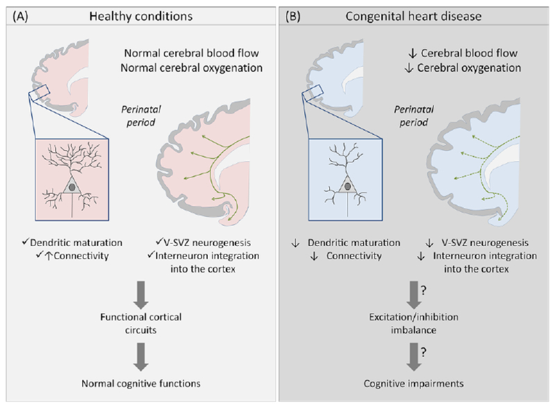
Congenital heart disease (CHD) is among the most common birth defects. Children with CHD frequently display long-term intellectual and behavioral disability. Emerging evidence indicates that cardiac anomalies lead to a reduction in cerebral oxygenation, which appears to profoundly impact on the maturation of cerebral regions responsible for higher-order cognitive functions. In this review we focus on the potential mechanisms by which dysregulation of cortical neuronal development during early life may lead to the significant cognitive impairments that commonly occur in children with CHD. Further understanding of the mechanisms underlying cortical dysmaturation due to CHD will be necessary to identify strategies for neonatal neuroprotection and for mitigating developmental delays in this patient population.

Keywords: congenital; cortex; heart; hypoxia; interneuron; perinatal.


Figures

Figure 1, Key Figure -
Impact of congenital heart disease on perinatal cortical maturation and subsequent cerebral impairments. Cartoon illustrating perinatal cortical development in healthy conditions (A) and in congenital heart disease (B). Before corrective surgery, CHD reduces cerebral blood flow and oxygenation. In animal models, hypoxemia is associated with a disruption of subplate neuron maturation and connectivity prenatally, and a decrease of V-SVZ neurogenesis and interneuron populations in the frontal cortex postnatally. These alterations could result in a cortical excitation/inhibition imbalance and potentially explain the cellular basis for the spectrum of cognitive impairments observed in CHD patients. Green lines and arrows represent streams of neuroblasts in the V-SVZ migrating to the olfactory bulb and frontal lobe.
Figure 2 -
Figure 2 -
Cellular impairments in the perinatal brain associated with hypoxia and CHD. Postnatal hypoxia in the piglet reduces the width of the SVZ (A,C; Scale bars, 500 mm) and the number of neuroblasts (DCX+) in the SVZ (B,D; Scale bars, 50 mm). Similarly, CHD reduces neuroblast numbers (DCX+) in the SVZ of human infants (E-H; Scale bars, 50 mm). Postnatal hypoxia in the piglet also reduces the number of DCX+ immature neurons (I,J; Scale bars, 50 mm) and NeuN+ mature neurons (K,L; Scale bars, 500 mm) in layers II/III of the frontal cortex. Prenatal hypoxia in the sheep results in a decrease in dendritic arborization of subplate neurons. 3D reconstructions of the soma and full basal and apical dendritic arbors (O,P) of representative sheep subplate neurons (M,N). Branch order rank is indicated by color: 1st order– bright yellow, 2nd order – white, 3rd order – purple/hot pink, 4th order – bright green, 5th order – cyan blue, 6th order – orange, 7th order – slate gray, 8th order – salmon pink, 9th order – forest green, and 10th order – bright blue. Adapted from Morton et al. [4] and McClendon et al. [75] with permission of the publishers.
Figure I for Box 1 -
Simplified systemic hemodynamics in (A) normal fetal circulation, (B) d-transposition of the great arteries (d-TGA) and (C) hypoplastic left heart syndrome (HLHS). Black arrows represent the blood flow. Yellow arrows represent the cerebral blood supply. The pulmonary circulation is not included because the blood flow to the lungs is highly limited due to elevated pulmonary vascular resistance and relatively low lung volume prior to birth. DA, ductus arteriosus; FO, foramen ovale; IVC, inferior vena cava; LA, left atrium; LV, left ventricle; PT, pulmonary trunk; RA, right atrium; RV, right ventricle; SVC, superior vena cava. Adapted from Sun et al. [51] with permission of the publishers.

References

    1. Passingham RE and Smaers JB (2014) Is the prefrontal cortex especially enlarged in the human brain allometric relations and remapping factors. Brain. Behav. Evol. 84, 156–66 - PubMed
    1. Southwell DG et al. (2014) Interneurons from Embryonic Development to Cell-Based Therapy. Science (80- ). 344, - PMC - PubMed
    1. Paredes MF et al. (2016) Extensive migration of young neurons into the infant human frontal lobe. Science 354, aaf7073. - PMC - PubMed
    1. Morton PD et al. (2017) Abnormal neurogenesis and cortical growth in congenital heart disease. Sci. Transl. Med. 9, eaah7029. - PMC - PubMed
    1. Dubois J and Dehaene-Lambertz G (2015) Fetal and Postnatal Development of the Cortex: MRI and Genetics. Brain Mapp. An Encycl. Ref. 2, 11–19

Publication types

MeSH terms