

Development and Patterning of the Cochlea: From Convergent Extension to Planar Polarity
- PMID: 30617059
- PMCID: PMC6938661
- DOI: 10.1101/cshperspect.a033266
Development and Patterning of the Cochlea: From Convergent Extension to Planar Polarity
Abstract
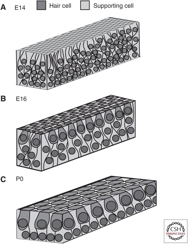
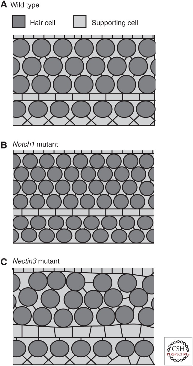
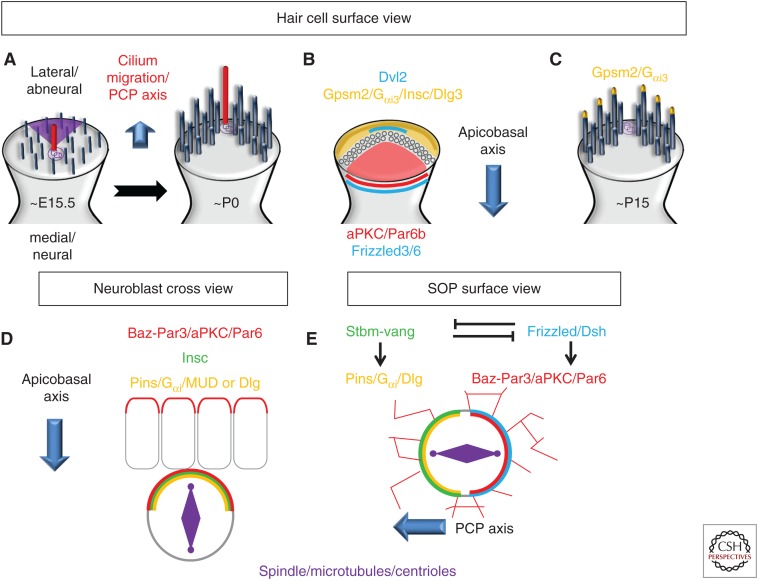
Within the mammalian cochlea, sensory hair cells and supporting cells are aligned in curvilinear rows that extend along the length of the tonotopic axis. In addition, all of the cells within the epithelium are uniformly polarized across the orthogonal neural-abneural axis. Finally, each hair cell is intrinsically polarized as revealed by the presence of an asymmetrically shaped and apically localized stereociliary bundle. It has been known for some time that many of the developmental processes that regulate these patterning events are mediated, to some extent, by the core planar cell polarity (PCP) pathway. This article will review more recent work demonstrating how components of the PCP pathway interact with cytoskeletal motor proteins to regulate cochlear outgrowth. Finally, a signaling pathway originally identified for its role in asymmetric cell divisions has recently been shown to mediate several aspects of intrinsic hair cell polarity, including kinocilia migration, bundle shape, and elongation.
Copyright © 2020 Cold Spring Harbor Laboratory Press; all rights reserved.
Figures
Similar articles
-
New insights into regulation and function of planar polarity in the inner ear.Neurosci Lett. 2019 Sep 14;709:134373. doi: 10.1016/j.neulet.2019.134373. Epub 2019 Jul 8. Neurosci Lett. 2019. PMID: 31295539 Free PMC article. Review.
-
Shaping the mammalian auditory sensory organ by the planar cell polarity pathway.Int J Dev Biol. 2007;51(6-7):535-47. doi: 10.1387/ijdb.072344mk. Int J Dev Biol. 2007. PMID: 17891715 Free PMC article. Review.
-
Wnt7b acts in concert with Wnt5a to regulate tissue elongation and planar cell polarity via noncanonical Wnt signaling.Proc Natl Acad Sci U S A. 2024 Aug 27;121(35):e2405217121. doi: 10.1073/pnas.2405217121. Epub 2024 Aug 22. Proc Natl Acad Sci U S A. 2024. PMID: 39172791 Free PMC article.
-
Evaluation of Planar-Cell-Polarity Phenotypes in Ciliopathy Mouse Mutant Cochlea.J Vis Exp. 2016 Feb 21;(108):53559. doi: 10.3791/53559. J Vis Exp. 2016. PMID: 26966880 Free PMC article.
-
Planar cell polarity and a potential role for a Wnt morphogen gradient in stereociliary bundle orientation in the mammalian inner ear.J Neurobiol. 2005 Sep 15;64(4):446-57. doi: 10.1002/neu.20171. J Neurobiol. 2005. PMID: 16041762 Review.
Cited by
-
The role of Rho GTPase family in cochlear hair cells and hearing.Neural Regen Res. 2023 Oct;18(10):2167-2172. doi: 10.4103/1673-5374.369101. Neural Regen Res. 2023. PMID: 37056125 Free PMC article. Review.
-
Vangl2 acts at the interface between actin and N-cadherin to modulate mammalian neuronal outgrowth.Elife. 2020 Jan 7;9:e51822. doi: 10.7554/eLife.51822. Elife. 2020. PMID: 31909712 Free PMC article.
-
A Reversal in Hair Cell Orientation Organizes Both the Auditory and Vestibular Organs.Front Neurosci. 2021 Sep 27;15:695914. doi: 10.3389/fnins.2021.695914. eCollection 2021. Front Neurosci. 2021. PMID: 34646115 Free PMC article. Review.
-
New insights into regulation and function of planar polarity in the inner ear.Neurosci Lett. 2019 Sep 14;709:134373. doi: 10.1016/j.neulet.2019.134373. Epub 2019 Jul 8. Neurosci Lett. 2019. PMID: 31295539 Free PMC article. Review.
-
Mapping Genome-wide Binding Sites of Prox1 in Mouse Cochlea Using the CUT&RUN Approach.Neurosci Bull. 2021 Dec;37(12):1703-1707. doi: 10.1007/s12264-021-00757-x. Epub 2021 Aug 5. Neurosci Bull. 2021. PMID: 34351548 Free PMC article. No abstract available.
References
-
- Aznar N, Ear J, Dunkel Y, Sun N, Satterfield K, He F, Kalogriopoulos NA, Lopez-Sanchez I, Ghassemian M, Sahoo D, Kufareva I, Ghosh P. 2018. Convergence of Wnt, growth factor, and heterotrimeric G protein signals on the guanine nucleotide exchange factor Daple. Sci Signal 11: eaao4220 10.1126/scisignal.aao4220 - DOI - PMC - PubMed
Publication types
MeSH terms
LinkOut - more resources
Full Text Sources