Review
. 2019 Jan 7;476(1):1-23.
doi: 10.1042/BCJ20180022.

Understanding phosphoinositides: rare, dynamic, and essential membrane phospholipids

Affiliations
Review

Understanding phosphoinositides: rare, dynamic, and essential membrane phospholipids

Eamonn J Dickson et al. Biochem J. .

Abstract

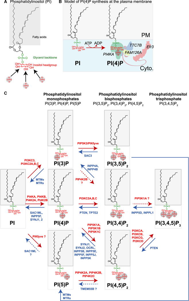
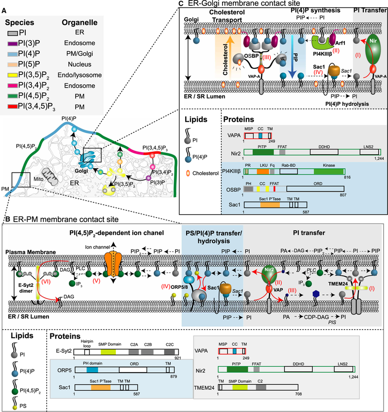
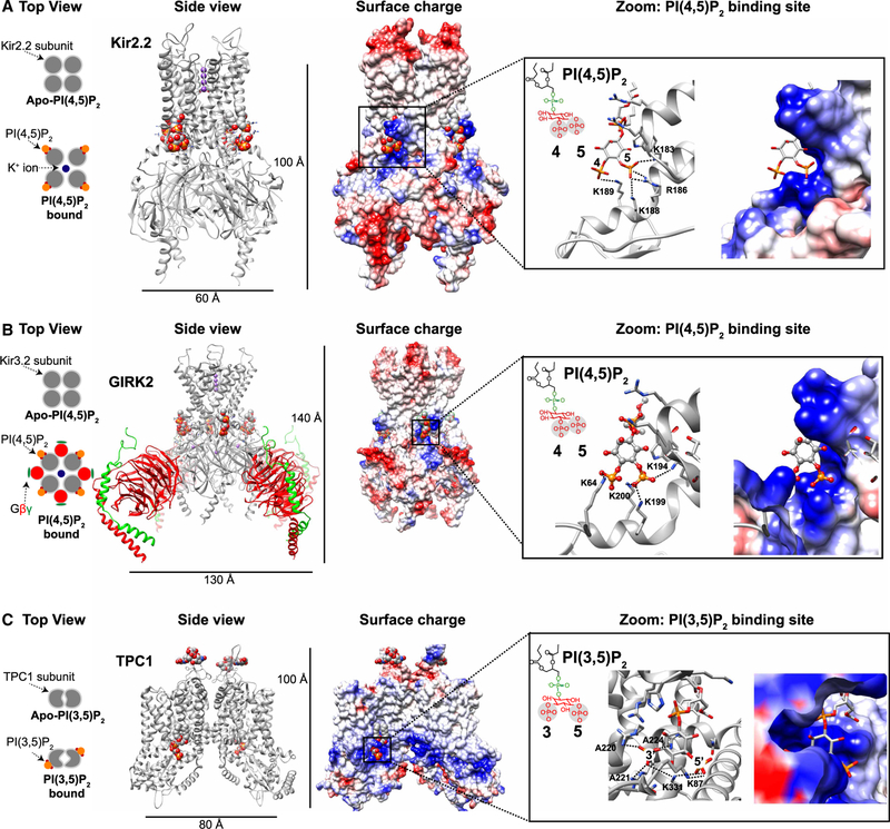
Polyphosphoinositides (PPIs) are essential phospholipids located in the cytoplasmic leaflet of eukaryotic cell membranes. Despite contributing only a small fraction to the bulk of cellular phospholipids, they make remarkable contributions to practically all aspects of a cell's life and death. They do so by recruiting cytoplasmic proteins/effectors or by interacting with cytoplasmic domains of membrane proteins at the membrane-cytoplasm interface to organize and mold organelle identity. The present study summarizes aspects of our current understanding concerning the metabolism, manipulation, measurement, and intimate roles these lipids play in regulating membrane homeostasis and vital cell signaling reactions in health and disease.

Keywords: PTEN; lipid transfer; phosphatidylinositol; phosphoinositide 3-kinase; transmembrane proteins.


Conflict of interest statement

Competing Interests

The Authors declare that there are no competing interests associated with the manuscript.

Figures

Figure 1.
Figure 1.. PPI biogenesis.
(A) Chemical structure of 1-stearoyl 2-arachidonoyl phosphatidylinositol (PI 38:4). Arrows pointing at the D3, D4, and D5 position on the myo-inositol head group indicate the three reversibly phosphorylatable hydroxyl groups. (B) Model of PI(4)P production from PI at the cytoplasmic (cyto.) interface of the plasma membrane (PM). (C) Diagram summarizing major PPI lipid kinase and phosphatase reaction pathways. Red arrows represent the PPI lipid kinases and blue arrows represent PPI lipid phosphatases. Red and blue labels are the gene name of enzymes capable of catalyzing each reaction. Gene names with question marks (?) and dashed reaction arrows represent enzymes with some ambiguity surrounding their ability to catalyze a specific reaction.
Figure 2.
Figure 2.. PPI metabolism at MCSs.
(A) Diagrammatic representation of the preferential distribution of PPIs in a eukaryotic cell. (B) Known proteins residing in ER– PM MCSs arranged in a hypothetical scenario. It remains to be fully tested whether each MCS contains this full standardized set of proteins or rather just a subset. For the dynamic equilibrium of PPI metabolism, (I) PI is transferred from the ER to the PM via TMEM24 dimers and this PI is then converted to PI(4)P by PM PI 4-kinases. (II) Under times of VAP-mediated Nir2 recruitment, PI would be transferred to the PM in exchange for phosphatidic acid (PA). (III) PA is subsequently converted to PI in a multi-step reaction. (IV) ORP5/8 countertransports PM PI(4)P to the ER in exchange for phosphatidylserine. Transferred PI (4)P is subsequently dephosphorylated to PI by the ER-resident phosphatase Sac1. (V) PM PI(4,5)P2 supports PM ion channel function and is also the substrate of PLC. Receptor-mediated activation of PLC hydrolyzes PM PI(4,5)P2 into IP3 and DAG. IP3 binds to IP3 receptors on ER membranes and to initiate release of Ca2+ from the ER, while DAG recruits PKC. Excess PM DAG may be cleared from the PM to the ER via extended synaptotagmin 2 (E-Syt2). (C) Known proteins residing in ER-Golgi MCSs. (I) PI is transferred from the ER to Golgi via VAP-A-Nir2 interactions. (II) Golgi PI is the substrate for PI4KIIIβ which generates PI (4)P in an Arf-1-dependent manner. (III) OSBP tethers ER-Golgi membranes through FFAT-mediated interactions with VAP-A on ER membranes and PH domain binding of PI(4)P on Golgi membranes. The ORD (OSBP-related domain) domain of OSBP can bind and transport PI(4)P from Golgi to ER membranes and cholesterol (against its concentration gradient) to Golgi membranes. (IV) Transferred ER PI(4)P is subsequently dephosphorylated into PI by ER Sac1, supporting the steep PI(4)P gradient between the two membranes.
Figure 3.
Figure 3.. Structural basis of PIP2 interactions with several ion channels.
In each case, the lipid is the diC8 version of a PIP2. (A) Top view: schematic representation of Kir2.2 subunits before and after PI(4,5)P2 binding. Side view: X-ray crystal structures of the Kir2.2 homotetrameric channel in presence of PI(4,5)P2 (gray, PDB: 3SPI [152]). Each channel subunit is in complex with a single PI(4,5)P2 molecule represented as spheres and colored according to atom type: carbon, gray; phosphorous, orange; and oxygen, red. Surface charge: electrostatic map of Kir2.2 homotetrameric channel structure (blue is positive; red negative; white neutral). The PI(4,5)P2 binding site comprises numerous basic residues (blue) that interact electrostatically with the negatively charged phosphates of PI(4,5)P2. Zoom: PI(4,5)P2 binding site. Note that positively charged residues in the protein structure are closely apposed to and predicted to interact with the negatively charged phosphate groups at the D4 and D5 positions of PI(4,5)P2. (B) Top view: schematic representation of Kir3.2 (GIRK2) subunits before and after PI(4,5)P2 binding. Side view: X-ray crystal structure of the Kir3.2 homotetrameric channel with accompanying Gβγ subunits (gray, PDB: 3SYA [153]). Surface charge: electrostatic map of Kir3.2 homotetrameric channel structure. Zoom: PI (4,5)P2 binding site. (C) Top view: schematic representation of TPC1 subunits before and after PI(3,5)P2 binding. Side view: cryo-EM structure of the endolysosomal TPC1 homodimeric channel in the PI(3,5)P2-bound state (gray, PDB: 6C9A [155]). Note that similar to Kir2.1 and Kir3.2, one PI(3,5)P2 molecule binds to each TPC1 subunit. Surface charge: electrostatic map of TPC1 homodimeric channel structure. Zoom: PI(3,5)P2 binding site.

Similar articles

Cited by

References

    1. Kim YJ, Guzman-Hernandez ML and Balla T (2011) A highly dynamic ER-derived phosphatidylinositol-synthesizing organelle supplies phosphoinositides to cellular membranes. Dev. Cell 21, 813–824 10.1016/j.devcel.2011.09.005 - DOI - PMC - PubMed
    1. Baskin JM, Wu X, Christiano R, Oh MS, Schauder CM, Gazzerro E et al. (2015) The leukodystrophy protein FAM126A (hyccin) regulates PtdIns(4)P synthesis at the plasma membrane. Nat. Cell Biol 18, 132–138 10.1038/ncb3271 - DOI - PMC - PubMed
    1. Nakatsu F, Baskin JM, Chung J, Tanner LB, Shui G, Lee SY et al. (2012) Ptdins4p synthesis by PI4KIIIα at the plasma membrane and its impact on plasma membrane identity. J. Cell Biol 199, 1003–1016 10.1083/jcb.201206095 - DOI - PMC - PubMed
    1. De Matteis M, Godi A and Corda D (2002) Phosphoinositides and the Golgi complex. Curr. Opin. Cell Biol 14, 434–447 10.1016/S0955-0674(02)00357-5 - DOI - PubMed
    1. Godi A, Pertile P, Meyers R, Marra P, Di Tullio G, Iurisci C et al. (1999) ARF mediates recruitment of PtdIns-4-OH kinase-beta and stimulates synthesis of PtdIns(4,5)P2 on the Golgi complex. Nat. Cell Biol 1, 280–287 10.1038/12993 - DOI - PubMed

Publication types