. 2019 Jan 8;3(1):33-46.
doi: 10.1182/bloodadvances.2018023804.

microRNA-22 promotes megakaryocyte differentiation through repression of its target, GFI1

Affiliations

microRNA-22 promotes megakaryocyte differentiation through repression of its target, GFI1

Cary N Weiss et al. Blood Adv. .

Abstract

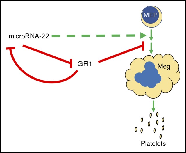
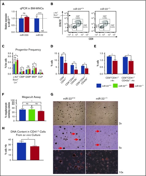
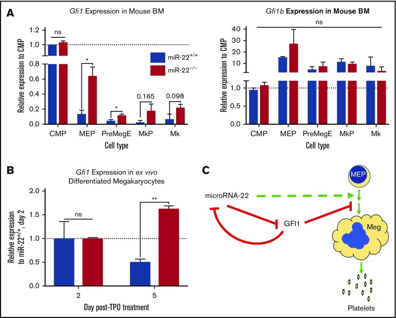
Precise control of microRNA expression contributes to development and the establishment of tissue identity, including in proper hematopoietic commitment and differentiation, whereas aberrant expression of various microRNAs has been implicated in malignant transformation. A small number of microRNAs are upregulated in megakaryocytes, among them is microRNA-22 (miR-22). Dysregulation of miR-22 leads to various hematologic malignancies and disorders, but its role in hematopoiesis is not yet well established. Here we show that upregulation of miR-22 is a critical step in megakaryocyte differentiation. Megakaryocytic differentiation in cell lines is promoted upon overexpression of miR-22, whereas differentiation is disrupted in CRISPR/Cas9-generated miR-22 knockout cell lines, confirming that miR-22 is an essential mediator of this process. RNA-sequencing reveals that miR-22 loss results in downregulation of megakaryocyte-associated genes. Mechanistically, we identify the repressive transcription factor, GFI1, as the direct target of miR-22, and upregulation of GFI1 in the absence of miR-22 inhibits megakaryocyte differentiation. Knocking down aberrant GFI1 expression restores megakaryocytic differentiation in miR-22 knockout cells. Furthermore, we have characterized hematopoiesis in miR-22 knockout animals and confirmed that megakaryocyte differentiation is similarly impaired in vivo and upon ex vivo megakaryocyte differentiation. Consistently, repression of Gfi1 is incomplete in the megakaryocyte lineage in miR-22 knockout mice and Gfi1 is aberrantly expressed upon forced megakaryocyte differentiation in explanted bone marrow from miR-22 knockout animals. This study identifies a positive role for miR-22 in hematopoiesis, specifically in promoting megakaryocyte differentiation through repression of GFI1, a target antagonistic to this process.


Conflict of interest statement

Conflict-of-interest disclosure: The authors declare no competing financial interests.

Figures

None
Graphical abstract
Figure 1.
Figure 1.
miR-22 expression increases upon terminal MK maturation and drives the maturation process. (A) Bone marrow from individual wildtype 129SV mice was stained according to surface markers listed in supplemental Table 3 and DAPI for live/dead assessment. Common myeloid progenitor (CMP), MEP, and MK were sorted by FACS for analysis of miR-22-3p expression by qPCR. sno202 was used as a housekeeping gene to quantify relative expression. Expression is shown relative to CMP, with the dotted line showing Relative Expression = 1. Data represent 2 independent experiments, performed in triplicate. (B) Representative qPCR for miR-22 in K562 cells treated with 15 nM PMA over time. K562 cells are driven to megakaryocytic differentiation. sno202 was used as a housekeeping gene to quantify relative expression. Expression is shown relative to K562 treated with vehicle (Veh; DMSO) at 24 hours. For the statistical analysis, the time points were treated as replicates. (C) miR-22 overexpression promotes MK maturation. K562 cells were transiently transfected with an miR-22 overexpression vector (PIG/miR-22) or a control (PIG/empty) and were subjected to PMA-driven megakaryocytic differentiation. GFP expression was used as a correlate for miR-22 overexpression and gated from I, no miR-22 overexpression, to IV, highest miR-22 overexpression (upper). Quantitation of percentages of CD61+ cells upon differentiation with 75 nM PMA or vehicle treatment and escalating empty vector or miR-22 expression (lower). (D) Utilizing CRISPR/Cas9 to knockout the miR-22 encoding stem loop from the MIR22HG in K562 cells. Schematic of the human MIR22HG on chromosome 17, specifically exon 2, which encodes the miR-22 stem loop (upper). Predicted schematics before and after locus excision and repair are shown, as well as gRNAs and genotyping primers. Agarose genotyping gel shows isolation of 6 clones each of scramble (ie, wildtype), miR-22 heterozygous, and miR-22 knockout (lower). Excision is identified by appearance of the truncated ∼480-bp band (external primers) and loss of the ∼360-bp band (internal primers). (E) qPCR for miR-22 expression in wildtype (ie, Scramble), miR-22 heterozygous, and miR-22 knockout K562 clones. sno202 was used as a housekeeping gene to quantify relative expression. Expression is shown relative to Scramble (n = 6). (F-G) K562:miR-22KO clones were subjected to PMA-driven megakaryocytic differentiation and assayed for differentiation by CD61 expression and nuclear content (ie, increased ploidy). (F) PMA-driven megakaryocytic differentiation over 48 hours in K562:miR-22KO clones was assessed by flow cytometry for CD61 expression and reported as fold change in median CD61 expression normalized per clone (n = 3). (G) PMA-driven megakaryocytic differentiation over 72 hours in K562:miR-22KO clones. Frequency of high-ploidy cells was assessed by flow cytometry. Gating strategy for identifying high-ploidy cells is shown (left) and is quantified in K562:Scramble and K562:miR-22KO clones (right) (n = 5). GFP, green fluorescent protein; SSC-A, side-scatter area. *P ≤ .05; **P < .01; ****P < .0001.
Figure 2.
Figure 2.
Identification of putative targets of miR-22 that may mediate its effect on megakaryopoiesis. (A) Venn diagram showing the strategy for selecting putative targets of miR-22 from RNA-sequencing data. K562:scramble and K562:miR-22KO were subjected to total RNA isolation, library preparation, and RNA-sequencing and analysis. To identify putative direct targets of miR-22, all targets needed to contain miR-22 seed sequences conserved in human and mouse; the yellow circle represents the top 30% of predicted TargetScan targets from the multimiR package (R). The blue circle represents genes upregulated in K562:miR-22KO cells, as would be expected of direct targets of miR-22. Four putative miR-22 targets were identified. The 2 chosen for further study are outlined in red. (B) Schematic showing the locations of predicted miR-22 seed sequences in human and mouse in the 3′-UTRs of the genes chosen for further study. (C-D) qPCR to confirm dysregulation of putative miR-22 targets chosen for future study in K562:miR-22KO cells (n = 3) (C). Expression is shown relative to scramble clone SCR3. (D) qPCR to confirm dysregulation of putative miR-22 targets chosen for future study miR-22 overexpressing cells. Expression is shown relative to p1501:Empty. GUSB was used as a housekeeping gene to quantify relative expression. **P < .01; ***P < .001. CDS, coding sequence.
Figure 3.
Figure 3.
Knockdown of miR-22 targets rescues the MK differentiation defect that results from miR-22 loss. (A-B) We used the CRISPRi (CRISPR interference) approach for knockdown of putative miR-22 target genes in K562:miR-22KO. K562:scramble and K562:miR-22KO cell lines were transduced with lentivirus encoding KRAB:dCas9-P2A-mCherry. The KRAB-dCas9 fusion protein is a strong transcriptional repressor that can be targeted by gRNAs to specific sites in the genome. A GFP targeting gRNA was used as a control. Putative miR-22 targets were knocked down by CRISPRi in K562:scramble and K562:miR-22KO cells, and cells were driven toward megakaryocytic differentiation by treatment with PMA. Extent of differentiation is quantified by DNA content/ploidy (n = 3) (A), and median CD61 expression (n = 5) (B). (C-E) Megakaryocytic differentiation of K562:scramble and K562:miR-22KO cells by treatment with PMA and assayed for transcript and protein expression. (C) qPCR for miR-22 and GFI1 upon PMA-induced megakaryocytic differentiation. sno202 and GUSB were used as housekeeping genes to quantify relative expression, respectively. Expression is shown relative to scramble clone SCR3 (n = 3). (D) Representative immunoblot against GFI1 in K562:miR-22KO cells upon PMA-induced megakaryocytic differentiation and in vehicle-treated control. Glyceraldehyde-3-phosphate dehydrogenase (GAPDH) was used as an endogenous loading control. (E) Quantitation of immunoblot against GFI1 in K562:miR-22KO cells upon PMA-induced megakaryocytic differentiation and in vehicle treated control. GAPDH was used as an endogenous loading control to quantitate relative expression. Quantified blot is included in supplemental Figure 3C (n = 3). (F) The miR-22 seed sequence containing portion of the GFI1 3′-UTR or a nontargeted control in which the seed sequence was replaced with poly-T tract was cloned downstream of the nanoLuciferase (nanoLuc) gene. The nanoLuc expression vectors were transiently cotransfected with a firefly luciferase expression vector into K562:scramble and K562:miR-22KO cells, and luminescence was quantified after 48 hours using the Nano-Glo Dual Luciferase Reporter Assay System. Quantitation is luminescence of nanoLuc (experimental vector) over luminescence of firefly luciferase (transfection control) (n = 3). (dT)7, poly-T tract; KO, knockout; SCR, scramble. *P ≤ .05; **P < .01.
Figure 4.
Figure 4.
miR-22 knockout mice exhibit an expansion of MK-erythrocyte progenitors and a defect in MK maturation. (A-C) Bone marrow from adult 129SV;miR-22 wildtype, heterozygous, and homozygous knockouts was isolated and subjected to analysis for gene expression, flow cytometric analysis, and ex vivo megakaryocytic differentiation. (A) Total RNA was isolated from bone marrow from wildtype and miR-22KO animals. RT was carried out using miRNA-specific primers. miR-223 was assayed as a control. sno202 was used as a housekeeping gene to quantify relative expression between samples (n = 3). (B) Bone marrow mononuclear cells from individual mice were stained for progenitors (CLP, CMP, GMP, and MEP) according to surface markers listed in supplemental Table 3, and DAPI for live/dead assessment. Shown are representative flow cytometry plots gated for myeloid progenitors from c-Kit+Sca1Lineage cells in wildtype and miR-22KO animals. (C) Quantitation of flow cytometric analysis of hematopoietic progenitor cells from wildtype, heterozygous, and miR-22KO animals (n = 6-9). (D-E) Bone marrow mononuclear cells from individual mice were stained for immature (CD9+CD41+CD42b) and mature (CD9+CD41+CD42b+) MKs, and for DNA content. Quantitation of frequency of immature and mature MKs (D), and quantitation of the frequency of high-ploidy cells in mature MKs (n = 3) (E). (F) CFU-MK assays. One thousand KSL were isolated from individual wildtype, heterozygous, and miR-22KO animals by FACS and were plated in 1.7 mL MegaCult supplemented with collagen and cytokines and plated in covered chamber slides for culture. After 7 days, cultures were dehydrated in acetone and stained for acetylcholinesterase and counterstained with Harris’ hematoxylin. CFU-MK and non-MK were quantified by a blinded counter on 2 separate days, and counts were averaged (n = 3). (G-H) Ex vivo MK differentiation of primary miR-22KO bone marrow cells. Bone marrow mononuclear cells were isolated from individual adult 129SV:miR-22 wildtype and miR-22KO and were subjected to ex vivo MK differentiation by treatment with TPO. Whole cultures were used for flow cytometry and acetylcholinesterase staining. (G) Representative microscopic images of acetylcholinesterase-stained cytospins from unfractionated ex vivo MK differentiation cultures. MKs are stained brown. Red arrows show MKs at 5× magnification. (H) Frequency of CD41+ cells with high DNA content (>4 n) in ex vivo differentiated MKs (n = 2-3). CFU-MK, colony forming unit–megakaryocyte; CLP, common lymphoid progenitor; CMP, common myeloid progenitor; GMP, granulocyte-monocyte progenitor; MEP, megakaryocyte-erythrocyte progenitor; nd, not detectable. *P ≤ .05; **P < .01; ****P < .0001.
Figure 5.
Figure 5.
Gfi1 expression persists in miR-22KOanimals throughout megakaryocytic differentiation. (A) Bone marrow from individual adult 129SV;miR-22 wildtype and miR-22 homozygous knockouts was stained according to surface markers listed in supplemental Table 3, DAPI for live/dead assessment, and sorted by FACS. Gene expression by qPCR for Gfi1 (left) and Gfi1b (right) in common myeloid progenitors through MK differentiation is shown. ActB was used as a housekeeping gene to quantify relative expression. Expression is shown relative to miR-22+/+ CMP, with the dotted line showing Relative Expression = 1 (n = 2-3). Error bars represent standard error of the mean. (B) Ex vivo MK differentiation of primary miR-22KO bone marrow cells. Bone marrow mononuclear cells were isolated from individual adult 129SV:miR-22 wildtype and miR-22KO and were subjected to ex vivo MK differentiation by treatment with TPO. For gene expression analysis, MKs were enriched by 2-step bovine serum albumin gradient sedimentation. qPCR for Gfi1 in ex vivo differentiated MKs at 2 and 5 days after initiation of TPO treatment. ActB was used as a housekeeping gene to quantify relative expression. Expression is shown relative to day 2 miR-22+/+ MKs, with the dotted line showing Relative Expression = 1 (n = 2-3). Error bars represent standard error of the mean. (C) Proposed model whereby repression of GFI1 by miR-22 permits megakaryopoiesis. miR-22 promotes megakaryopoiesis (dotted green line) through direct repression of GFI1 (solid red line). GFI1 represses miR-22 expression by binding at the promoter of the MIR22HG. BM, bone marrow; MkP, megakaryocyte progenitor; PreMegE, megakaryocyte-erythrocyte precursor. *P ≤ .05; **P < .01.

Similar articles

Cited by

References

    1. Bluteau D, Lordier L, Di Stefano A, et al. . Regulation of megakaryocyte maturation and platelet formation. J Thromb Haemost. 2009;7(Suppl 1):227-234. - PubMed
    1. Kaushansky K. Historical review: megakaryopoiesis and thrombopoiesis. Blood. 2008;111(3):981-986. - PMC - PubMed
    1. Geddis AE. Megakaryopoiesis. Semin Hematol. 2010;47(3):212-219. - PMC - PubMed
    1. Pinho S, Marchand T, Yang E, Wei Q, Nerlov C, Frenette PS. Lineage-biased hematopoietic stem cells are regulated by distinct niches. Dev Cell. 2018;44(5):634-641.e634. - PMC - PubMed
    1. Boulais PE, Frenette PS. Making sense of hematopoietic stem cell niches. Blood. 2015;125(17):2621-2629. - PMC - PubMed

Publication types