

Cell migration: implications for repair and regeneration in joint disease
- PMID: 30617265
- PMCID: PMC7004411
- DOI: 10.1038/s41584-018-0151-0
Cell migration: implications for repair and regeneration in joint disease
Abstract
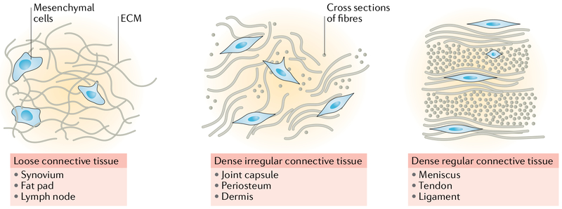
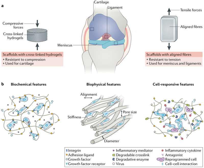
Connective tissues within the synovial joints are characterized by their dense extracellular matrix and sparse cellularity. With injury or disease, however, tissues commonly experience an influx of cells owing to proliferation and migration of endogenous mesenchymal cell populations, as well as invasion of the tissue by other cell types, including immune cells. Although this process is critical for successful wound healing, aberrant immune-mediated cell infiltration can lead to pathological inflammation of the joint. Importantly, cells of mesenchymal or haematopoietic origin use distinct modes of migration and thus might respond differently to similar biological cues and microenvironments. Furthermore, cell migration in the physiological microenvironment of musculoskeletal tissues differs considerably from migration in vitro. This Review addresses the complexities of cell migration in fibrous connective tissues from three separate but interdependent perspectives: physiology (including the cellular and extracellular factors affecting 3D cell migration), pathophysiology (cell migration in the context of synovial joint autoimmune disease and injury) and tissue engineering (cell migration in engineered biomaterials). Improved understanding of the fundamental mechanisms governing interstitial cell migration might lead to interventions that stop invasion processes that culminate in deleterious outcomes and/or that expedite migration to direct endogenous cell-mediated repair and regeneration of joint tissues.
Conflict of interest statement
Competing interests
The authors declare no competing interests.
Figures

Similar articles
-
The matrix environmental and cell mechanical properties regulate cell migration and contribute to the invasive phenotype of cancer cells.Rep Prog Phys. 2019 Jun;82(6):064602. doi: 10.1088/1361-6633/ab1628. Epub 2019 Apr 4. Rep Prog Phys. 2019. PMID: 30947151 Review.
-
Different wound healing properties of dermis, adipose, and gingiva mesenchymal stromal cells.Wound Repair Regen. 2016 Jan-Feb;24(1):100-9. doi: 10.1111/wrr.12380. Epub 2015 Dec 1. Wound Repair Regen. 2016. PMID: 26542883
-
Computational modelling of cell spreading and tissue regeneration in porous scaffolds.Biomaterials. 2007 Apr;28(10):1926-40. doi: 10.1016/j.biomaterials.2006.12.008. Epub 2006 Dec 18. Biomaterials. 2007. PMID: 17178156 Review.
-
Plasticity in cell migration modes across development, physiology, and disease.Front Cell Dev Biol. 2024 Apr 23;12:1363361. doi: 10.3389/fcell.2024.1363361. eCollection 2024. Front Cell Dev Biol. 2024. PMID: 38715921 Free PMC article. Review.
-
Programmed biomolecule delivery to enable and direct cell migration for connective tissue repair.Nat Commun. 2017 Nov 24;8(1):1780. doi: 10.1038/s41467-017-01955-w. Nat Commun. 2017. PMID: 29176654 Free PMC article.
Cited by
-
In vivo bone regeneration assessment of offset and gradient melt electrowritten (MEW) PCL scaffolds.Biomater Res. 2020 Oct 1;24:17. doi: 10.1186/s40824-020-00196-1. eCollection 2020. Biomater Res. 2020. PMID: 33014414 Free PMC article.
-
In Vitro Comparison of 2 Clinically Applied Biomaterials for Autologous Chondrocyte Implantation: Injectable Hydrogel Versus Collagen Scaffold.Cartilage. 2023 Jun;14(2):220-234. doi: 10.1177/19476035231154507. Epub 2023 Mar 1. Cartilage. 2023. PMID: 36859785 Free PMC article.
-
Preliminary Exploration of Al18F-NOTA-FAPI-04 PET/CT in the Management of Ankylosing Spondylitis: A Prospective Clinical Study.Mol Imaging. 2024 Sep 8;23:15353508241270405. doi: 10.1177/15353508241270405. eCollection 2024 Jan-Dec. Mol Imaging. 2024. PMID: 40230596 Free PMC article.
-
Comparative analysis of extracellular vesicles from induced and adipose-derived Mesenchymal Stem Cells: Implications for regenerative medicine.PLoS One. 2025 Jun 4;20(6):e0325065. doi: 10.1371/journal.pone.0325065. eCollection 2025. PLoS One. 2025. PMID: 40465768 Free PMC article.
-
Maneuvering the Migration and Differentiation of Stem Cells with Electrospun Nanofibers.Adv Sci (Weinh). 2020 Jun 9;7(15):2000735. doi: 10.1002/advs.202000735. eCollection 2020 Aug. Adv Sci (Weinh). 2020. PMID: 32775158 Free PMC article. Review.
References
-
- Franz CM, Jones GE & Ridley AJ Cell migration in development and disease. Dev. Cell 2, 153–158 (2002). - PubMed
-
- Cukierman E, Pankov R, Stevens DR & Yamada KM Taking cell-matrix adhesions to the third dimension. Science 294, 1708–1712 (2001). - PubMed
-
- Pathak A & Kumar S Biophysical regulation of tumor cell invasion: moving beyond matrix stiffness. Integr. Biol 3, 267–278 (2011). - PubMed
Publication types
MeSH terms
Grants and funding
LinkOut - more resources
Full Text Sources
Medical
