

Ocular abnormalities and complications in anterior megalophthalmos: a case series
- PMID: 30617289
- PMCID: PMC6707321
- DOI: 10.1038/s41433-018-0329-3
Ocular abnormalities and complications in anterior megalophthalmos: a case series
Abstract
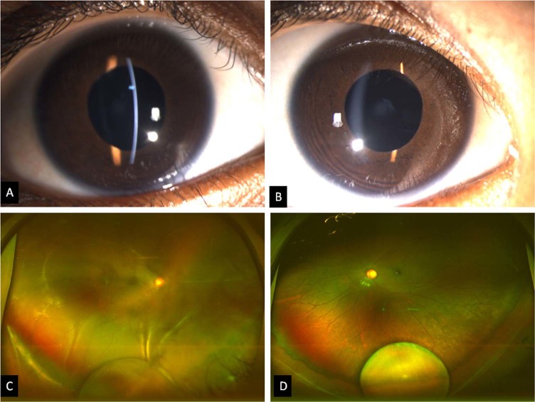
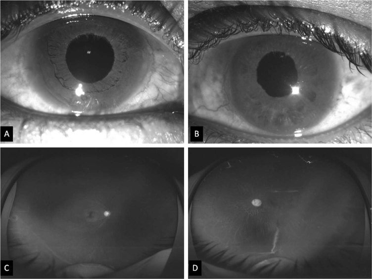
Objectives: To describe the clinical and Scheimpflug imaging features in cases of anterior megalophthalmos (AM).
Methods: Retrospective record review was performed for patients with AM who presented between June 2017 and May 2018. Clinical history, slit lamp examination, Scheimpflug imaging indices (Pentacam-HR, Oculus, GmbH), dilated fundus examination and treatment records were reviewed.
Results: The study included eight eyes of four male patients (mean age 6.5 years, range 4-10 years). Corrected distance visual acuity ranged from finger counting to 20/80. The mean horizontal corneal diameter, central corneal thickness, steep keratometry, flat keratometry, anterior chamber (AC) angle, AC depth, maximum pupil diameter and axial length were 13.8 ± 0.5 mm, 538.7 ± 68.9 µm, 42.8 ± 1.6 D, 41.4 ± 0.9D, 47.0 ± 4.2 degree, 3.8 ± 0.3 mm, 3.9 ± 0.1 mm, and 24.9 ± 0.9 mm, respectively. Posterior dislocation of crystalline lens, vitreous degeneration and rhegmatogenous retinal detachment (consequent of retinal dialysis/atrophic hole/lattice with hole) were noted in seven, eight and five eyes, respectively. Pigment dispersion glaucoma was noted in both eyes of one patient, which was managed with topical anti-glaucoma medication. Vitrectomy with silicone oil tamponade was successful in retinal reattachment in all three eyes that underwent surgery.
Conclusion: Scheimpflug imaging helps in demonstrating the corneal and anterior segment pathological changes in AM. The disease extends to involve the zonules, vitreous and retina as well. Ophthalmologists should be able to identify this disorder, recognise and manage the associations and complications.
Conflict of interest statement
The authors declare that they have no conflict of interest.
Figures
References
-
- Vail DT. Adult hereditary anterior megalophthalmus sine glaucoma: a definite disease entity: with special reference to the extraction of cataract. Arch Ophthalmol. 1931;6:39–62. doi: 10.1001/archopht.1931.00820070042004. - DOI
