. 2019 Mar;12(2):545-554.
doi: 10.1038/s41385-018-0115-3. Epub 2019 Jan 7.

Excessive neutrophil levels in the lung underlie the age-associated increase in influenza mortality

Affiliations

Excessive neutrophil levels in the lung underlie the age-associated increase in influenza mortality

Upasana Kulkarni et al. Mucosal Immunol. 2019 Mar.

Abstract

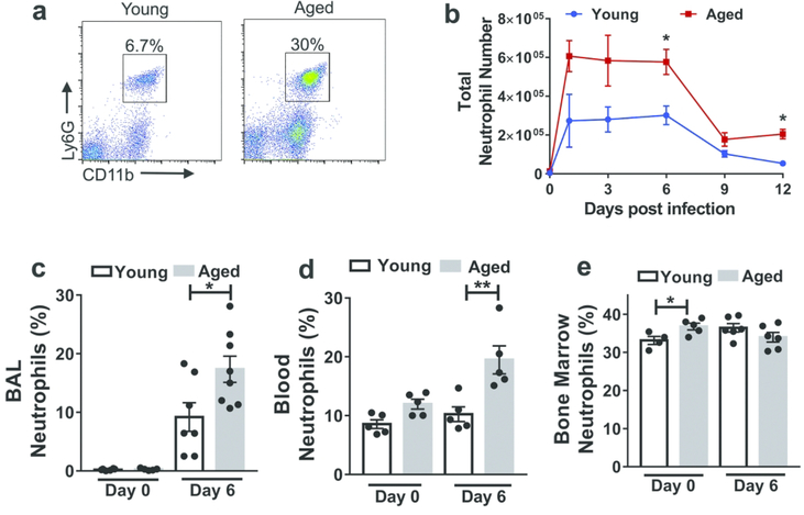
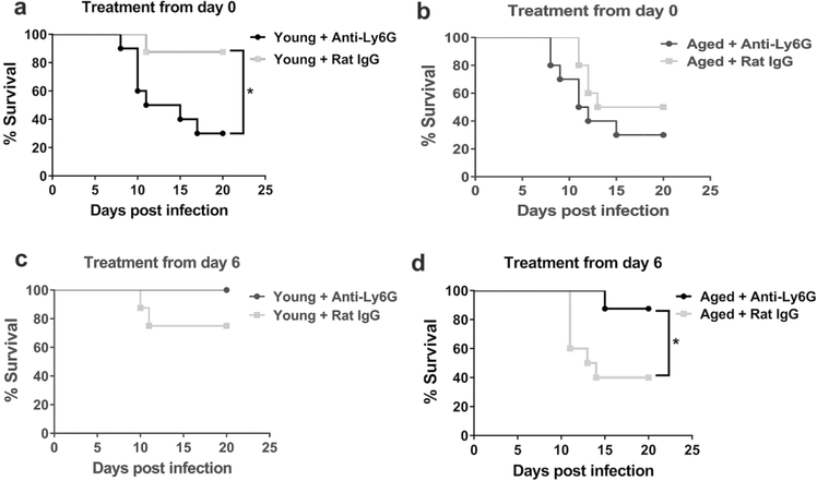
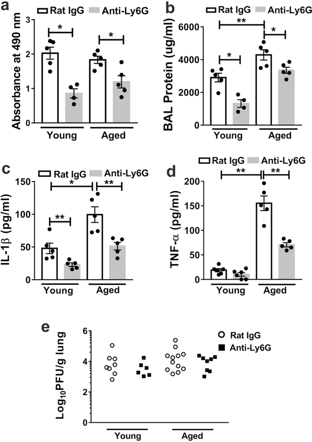
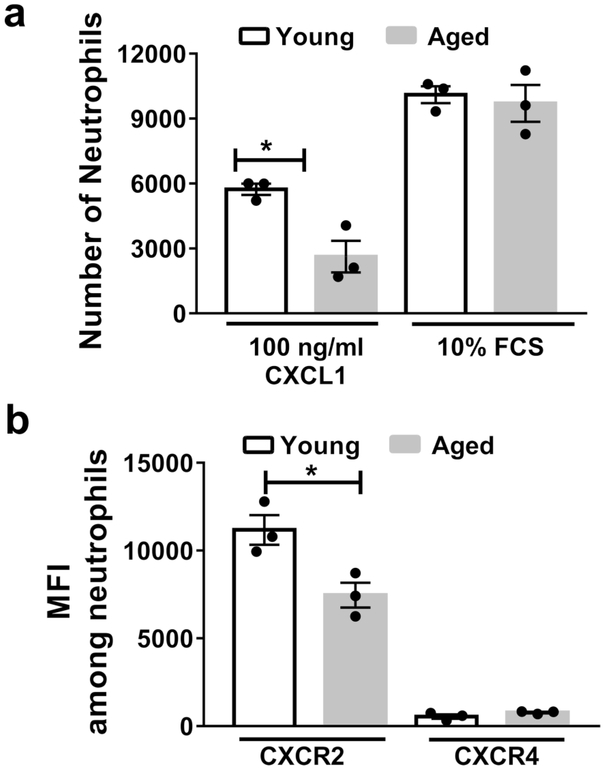
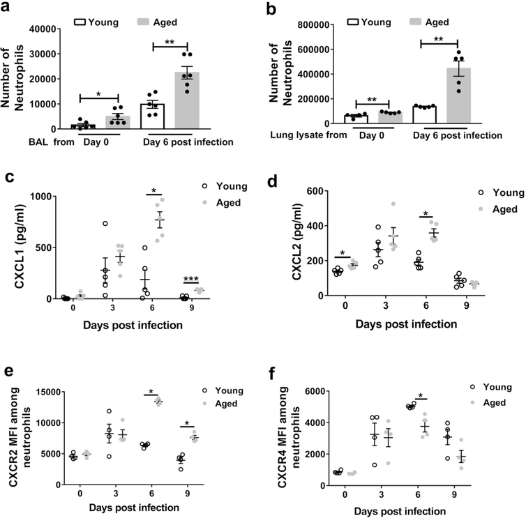
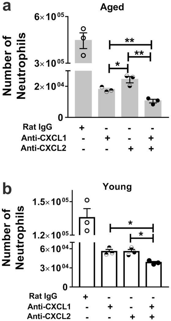
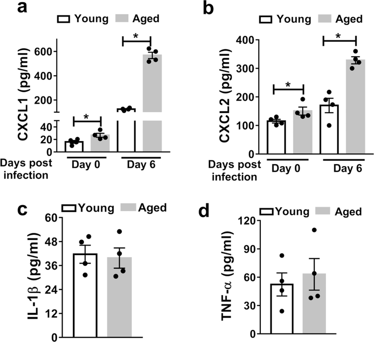
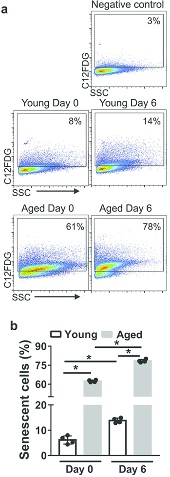
Neutrophils clear viruses, but excessive neutrophil responses induce tissue injury and worsen disease. Aging increases mortality to influenza infection; however, whether this is due to impaired viral clearance or a pathological host immune response is unknown. Here we show that aged mice have higher levels of lung neutrophils than younger mice after influenza viral infection. Depleting neutrophils after, but not before, infection substantially improves the survival of aged mice without altering viral clearance. Aged alveolar epithelial cells (AECs) have a higher frequency of senescence and secrete higher levels of the neutrophil-attracting chemokines CXCL1 and CXCL2 during influenza infection. These chemokines are required for age-enhanced neutrophil chemotaxis in vitro. Our work suggests that aging increases mortality from influenza in part because senescent AECs secrete more chemokines, leading to excessive neutrophil recruitment. Therapies that mitigate this pathological immune response in the elderly might improve outcomes of influenza and other respiratory infections.


Conflict of interest statement

Disclosure

The authors have declared that no conflict of interest exists.

Figures

Figure 1.
Figure 1.. Aging is associated with increased neutrophils within the lung during influenza infection.
Aged (18–22 months of age) and young (2–4 months of age) C57BL/6 mice were intranasally inoculated with PR8 strain influenza virus. Lung tissue was harvested, digested and cells were obtained and suspended to permit incubation with fluorescent monoclonal antibodies followed by flow cytometric analysis. a Representative flow cytometric plot at day 6 p.i. (post-infection) of a young and aged mouse showing increased proportion of neutrophils (CD11b+, Ly6Ghi) within the lung in the aged mouse than the young mouse. The flow cytometric plots are gated on CD11b+Ly6Ghi cells. b Absolute number of neutrophils before and during influenza infection in young and aged mice in entire lung (airspace, vasculature and lung interstitium). * P < 0.05, (Mann-Whitney test). n = 3–6 / group/ time point. Data representative of one of two independent experiments. c,d,e Proportion of neutrophils analyzed by flow cytometry in non-infected and infected (day +6 p.i.) young and aged mice in BAL (c) blood (d) and bone marrow (e). * P < 0.05, ** P < 0.01 (Mann-Whitney test). Data representative of one of two independent experiments,which yielded similar results.
Figure 2.
Figure 2.. Neutrophil depletion at the end of the first week p.i. (post-infection) enhances survival in aged mice.
a Young mice were administered a neutrophil depleting monoclonal antibody (i.p.) just prior to inoculation with influenza virus and following infection (as described in methods) and survival was monitored. Compared to isotype control treated, infected neutrophil depleted young mice exhibited a significant increase in mortality. * P < 0.05 (Log rank), n= 8–10 per group. b Aged mice were treated per A and survival post infection was monitored. P value between neutrophil depleted and control group P = 0.23 (Log-Rank). n= 8–10 per group. c Young mice were administered a neutrophil depleting antibody at day + 6 p.i. then intermittently to day +12 p.i. (as described in Methods) and compared to isotype control, treated young-infected mice. * P = 0.10 (Log-rank), n= 8–10 per group. d Aged mice were treated according to c and survival was measured post infection. Aged neutrophil, depleted mice exhibited a significant increased survival as compared to control group. * P < 0.05 Log-rank, n= 8–10 per group.
Figure 3.
Figure 3.. Neutrophil depletion in mice from the first week p.i. (post-infection) reduces lung damage and lung inflammatory cytokines levels without impacting viral clearance.
Aged and young mice were infected with influenza virus and depleted of neutrophils or treated with isotype control at day 6 p.i and then intermittently until day 12 p.i. At day 10 p.i., the BAL was obtained from and LDH (a), protein (b), IL-1β (c), TNF-α (d) measured via ELISA * P < 0.05 ** P = 0.01 (Mann-Whitney test). Data are representative of one of two independent experiments, which yielded similar results. Data are expressed as mean ± SEM eYoung and aged mice were infected with influenza and depleted of neutrophils or treated with isotype control at day 6 p.i and then intermittently until day 10 p.i. At day 10 p.i, lungs were obtained and viral load measured by plaque assay. There were no significant differences in viral load between control and neutrophil depleted groups. P = 0.49 (young Rat IgG vs Anti-Ly6G(Mann-Whitney test) and P = 0.47 (aged Rat IgG vs Anti-Ly6G) (Mann-Whitney test). Data are representative of two independent experiments.
Figure 4.
Figure 4.. Aging impairs bone marrow neutrophil chemotaxis towards CXCL1 and reduces surface expression of CXCR2.
Neutrophils were purified, via negative magnetic separation from the bone marrow of young and aged non-infected mice. a Neutrophil chemotaxis was assessed in response to CXCL1 or fetal calf serum (FCS) at the indicated doses. b Surface expression of CXCR2 and CXCR4 via flow cytometry was assessed on purified bone marrow neutrophils from young and aged mice. Experiments are representative of one experiment repeated twice with consistent results. * P< 0.05 (Mann-Whitney test). Data are expressed as mean ± SEM.
Figure 5.
Figure 5.. Aging increases the levels of CXCL1 and CXCL2 in lung during influenza infection and enhances neutrophil chemotaxis in vitro.
a-b Bone marrow neutrophils were purified from young non-infected mice and added to chemotaxis assays in which the chemoattractant was either the BAL (a) or lung lysate (b) of non-infected or infected (day 6 post-infection), young and aged mice. Young neutrophils exhibited a significant increased chemotaxis towards the fluid obtained from the lungs of aged mice than young mice (before or after infection) * P < 0.05 ** P < 0.01 (Mann-Whitney test). c-d BAL was obtained during the course of influenza infection in young and aged mice and CXCL1 (c) and CXCL2 (d) measured within the BAL via ELISA. n = 5/ time point / group. * P < 0.05 *** P < 0.001 (Mann-Whitney test). e-f Surface expression of CXCR2 and CXCR4 on neutrophils in the lung before and during the course of influenza infection in young and aged mice. n = 4/ time point / group. * P < 0.05 (Mann-Whitney test).
Figure 6.
Figure 6.. CXCL1 and CXCL2 are critical for chemotaxis of the influenza-infected lung with aging.
The lung lysate from young and aged, infected mice were obtained and employed in chemotaxis assays with neutrophils purified from the bone marrow of young mice. Inhibiting CXCL1 or CXCL2 reduced neutrophil chemotaxis of aged (a) and young (b) lung lysate (P < 0.05 vs. control in age group). n = 3–6. * P < 0.05 (Mann-Whitney test). Isotype control antibody did not significantly reduce neutrophil chemotaxis. Data representative of one of two independent experiments, which yielded similar results. Data are expressed as mean ± SEM.
Figure 7.
Figure 7.. AECs secrete higher levels of CXCL1 and CXCL2 with aging during influenza infection.
AECs were purified from young and aged non-infected and infected mice (six days post-infection) andcultured ex vivo. After 36h the supernatants were harvested and CXCL1 (a), CXCL2 (b) TNF-α (c) and IL-1β (d) were measured via ELISA. * P < 0.05 (Mann-Whitney test). Data are representative o ftwo independent experiments, repeated with similar results. Data are expressed as mean ± SEM.
Figure 8.
Figure 8.. AECs from aged mice upregulate senescence-associated β-galactosidase activity.
AECs were purified from young and aged non-infected and infected mice (six days post-infection) and cultured ex vivo. After 3 h, cells were incubated for 2h with C12FDG, a β-galactosidase substrate that produces fluorescent product upon cleavage. The β-galactosidase activity was measured via flow cytometry. Cells incubated with PBS were used as negative control. a Representative flow cytometry plots gated on cells expressing the cleaved fluorescent product.b Graph indicating the frequency of senescent cells showing β-galactosidase activity. * P<0.05

References

    1. Thompson WW, Shay DK, Weintraub E, Brammer L, Cox N, Anderson LJ et al. Mortality Associated With Influenza and Respiratory Syncytial Virus in the United States. JAMA: The Journal of the American Medical Association 2003; 289(2): 179–186. - PubMed
    1. Pebody RG, McLean E, Zhao H, Cleary P, Bracebridge S, Foster K et al. Pandemic Influenza A (H1N1) 2009 and mortality in the United Kingdom: risk factors for death, April 2009 to March 2010. Eurosurveillance 2010; 15(20): 19571. - PubMed
    1. Fry AM, Shay DK, Holman RC, Curns AT, Anderson LJ. Trends in Hospitalizations for Pneumonia Among Persons Aged 65 Years or Older in the United States, 1988–2002. JAMA 2005; 294(21): 2712–2719. - PubMed
    1. Biggerstaff M, Kniss K, Jernigan DB, Brammer L, Bresee J, Garg S et al. Systematic Assessment of Multiple Routine and Near Real-Time Indicators to Classify the Severity of Influenza Seasons and Pandemics in the United States, 2003–2004 Through 2015–2016. American Journal of Epidemiology 2018; 187(5): 1040–1050. - PMC - PubMed
    1. Shaw AC, Goldstein DR, Montgomery RR. Age-dependent dysregulation of innate immunity. Nat Rev Immunol 2013; 13(12): 875–887. - PMC - PubMed

Publication types