Review
. 2019 Apr;33(2):221-230.
doi: 10.1007/s10557-018-6845-3.

Cardiac Myosin-Binding Protein C-From Bench to Improved Diagnosis of Acute Myocardial Infarction

Affiliations
Review

Cardiac Myosin-Binding Protein C-From Bench to Improved Diagnosis of Acute Myocardial Infarction

Thomas E Kaier et al. Cardiovasc Drugs Ther. 2019 Apr.

Abstract

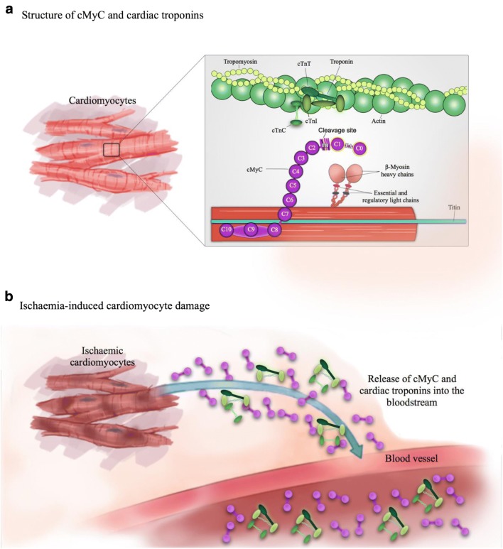
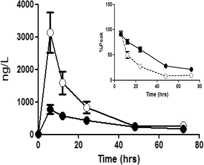
Chest pain is responsible for 6-10% of all presentations to acute healthcare providers. Triage is inherently difficult and heavily reliant on the quantification of cardiac Troponin (cTn), as a minority of patients with an ultimate diagnosis of acute myocardial infarction (AMI) present with clear diagnostic features such as ST-elevation on the electrocardiogram. Owing to slow release and disappearance of cTn, many patients require repeat blood testing or present with stable but elevated concentrations of the best available biomarker and are thus caught at the interplay of sensitivity and specificity.We identified cardiac myosin-binding protein C (cMyC) in coronary venous effluent and developed a high-sensitivity assay by producing an array of monoclonal antibodies and choosing an ideal pair based on affinity and epitope maps. Compared to high-sensitivity cardiac Troponin (hs-cTn), we demonstrated that cMyC appears earlier and rises faster following myocardial necrosis. In this review, we discuss discovery and structure of cMyC, as well as the migration from a comparably insensitive to a high-sensitivity assay facilitating first clinical studies. This assay was subsequently used to describe relative abundance of the protein, compare sensitivity to two high-sensitivity cTn assays and test diagnostic performance in over 1900 patients presenting with chest pain and suspected AMI. A standout feature was cMyC's ability to more effectively triage patients. This distinction is likely related to the documented greater abundance and more rapid release profile, which could significantly improve the early triage of patients with suspected AMI.

Keywords: AMI; Acute myocardial infarction; Biomarkers; Cardiac myosin-binding protein C; Cardiac troponin; Chest pain; Triage; cMyC.


Conflict of interest statement

Conflict of Interest

T.E.K. and B.A. have received research grants from the British Heart Foundation. T.E.K. has received speaker fees from Astra Zeneca. M.M. is named as an inventor on a patent held by King’s College London for the detection of cardiac myosin–binding protein C as a biomarker of myocardial injury.

Ethical Approval

All applicable international, national and/or institutional guidelines for the care and use of animals were followed. All procedures performed in studies involving human participants were in accordance with the ethical standards of the institutional and/or national research committee and with the 1964 Helsinki declaration and its later amendments or comparable ethical standards.

Figures

Fig. 1
Fig. 1
Structure of cMyC and relationship with the cTn complex; adapted from Kaier et al. [27]
Fig. 2
Fig. 2
Structure of full-length cMyBP-C. Phosphorylation sites involved in the regulation of myocardial contractility—Ser-273, Ser-282 and Ser-302—highlighted in the M-domain (where calpain-dependent cleavage occurs) [55], and commonly detected N-terminal fragments, C0C2 and C0C1f. Binding sites for antibodies 1A4 (blue) and 3H8 (red) are highlighted. Reproduced and adapted from Lipps et al. [56]
Fig. 3
Fig. 3
The development of a quantitative immunoassay for human cMyC in serum. a Sequence alignment of cMyC with skeletal myosin binding protein C isoforms. The sequence recognised by monoclonal anti-cMyC antibodies 1A4 and 3H8 are shown in bold. The antibodies bind to cardiac-restricted sequences with organ specificity further verified by immunoblots (see d). b SPR kinetic sensorgrams demonstrating the kinetic parameters of clone 3H8 (left) and 1A4 (right). These antibodies were selected from over 50 hybridomas, and both antibodies are of high affinity. c Epitope competition sensorgram of 1A4 and 3H8 binding to the C0C2 region of cMyC conjugated to a CM5 biosensor chip. Although antibodies recognise near adjacent epitopes, there is no appreciable interference between them. Near adjacency is needed since cMyC is fragmented in the circulation raising the possibility of separation of capture and detection epitopes if they were widely spaced. d Immunoblot of rat and human tissue demonstrating specificity of 3H8 and 1A4 monoclonal antibodies. GAPDH was used as a loading control. Samples 1–9 are various rat tissue (1 = ventricle, 2 = atria, 3 = rectus abdominus, 4 = soleus, 5 = spleen, 6 = kidney, 7 = aorta, 8 = liver, 9 = brain) and 10 is human ventricle. e Representative C0C2 standard curve from cMyC ECL assay indicating the limit of detection (dashed line). This in-house assay on a MesoScale Discovery enhanced chemiluminescent detection platform was used to measure cMyC appearance and disappearance in Figs. 2 and 3 below. Panel (f) demonstrates the performance characteristics of the assay, with a LoD of approximately 80 ng/L. Figures and legend reproduced from Baker et al. [22]
Fig. 4
Fig. 4
The accumulation of cMyC vs. cTnT after myocardial injury caused by intracoronary ethanol. Venous blood was collected frequently over the first 2 h, and up to 24 h, after therapeutic alcohol septal ablation for hypertrophic cardiomyopathy (TASH) using ethanol infused selectively into a septal perforating branch coronary artery. Summary data of absolute quantification of cMyC (open symbols) vs. cTnT (closed symbols) over time following TASH (n = 20). Inset figure is a zoom of the first 240 min. Over this time interval, cMyC accumulates in the serum approximately six times faster than cTnT (slope 25.8 ± 1.9 vs. 4.0 ± 0.4 ng/L/min, p < 0.0001). Figure reproduced from Baker et al. [22]
Fig. 5
Fig. 5
The accumulation of cMyC (open symbols) vs. cTnT (closed symbols) after myocardial injury caused by surgical revascularisation. Venous blood was collected over 3 days following CABG. Summary data of absolute quantification of cMyC vs. cTnT over time following CABG (n = 20). Inset figure is a zoom of the last five time points expressed as a % of peak concentration achieved in each patient. This normalisation was used to remove the visual bias caused by the greater absolute concentration of cMyC. The decay half-time for cMyC is considerably shorter than for cTnT (5.5 ± 0.8 h vs. 22 ± 5 h, p < 0.0001). Figure reproduced from Baker et al. [22]
Fig. 6
Fig. 6
Amino acid sequence of cMyBP-C with variant MET-158 underlined; antibodies 1A4 (blue) and 3H8 (red) at binding location

References

    1. Goodacre S. The health care burden of acute chest pain. Heart. 2005;91(2):229–230. - PMC - PubMed
    1. Murphy NF. Hospital discharge rates for suspected acute coronary syndromes between 1990 and 2000: population based analysis. BMJ. 2004;328(7453):1413–1414. - PMC - PubMed
    1. Blatchford O, Capewell S, Murray S, Blatchford M. Emergency medical admissions in Glasgow: general practices vary despite adjustment for age, sex, and deprivation. Br J Gen Pract. 1999;49(444):551–554. - PMC - PubMed
    1. Harris T, McDonald K. Is the case-mix of patients who self-present to ED similar to general practice and other acute-care facilities? Emerg Med J. 2014;31(12):970–974. - PubMed
    1. NHS Digital. Hospital Episode Statistics [Internet]. [cited 2018 Apr 30]. Available from: https://digital.nhs.uk/data-and-information/data-tools-and-services/data...