. 2018 Dec 20:12:970.
doi: 10.3389/fnins.2018.00970. eCollection 2018.

Retinal Neuroprotection From Optic Nerve Trauma by Deletion of Arginase 2

Affiliations

Retinal Neuroprotection From Optic Nerve Trauma by Deletion of Arginase 2

Zhimin Xu et al. Front Neurosci. .

Abstract

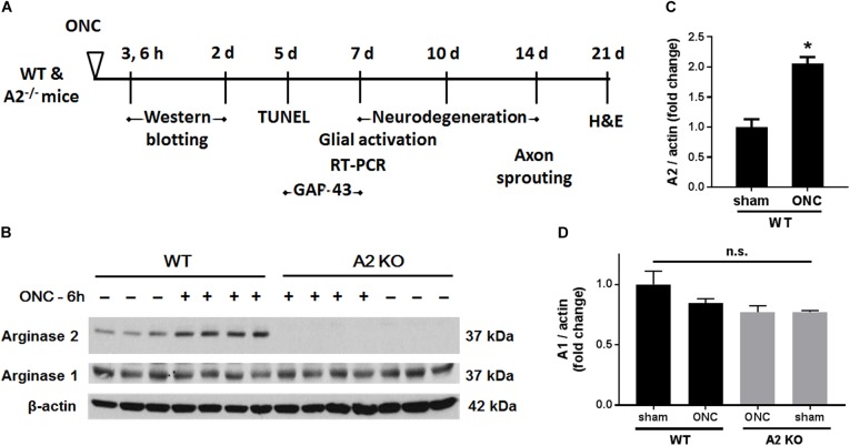
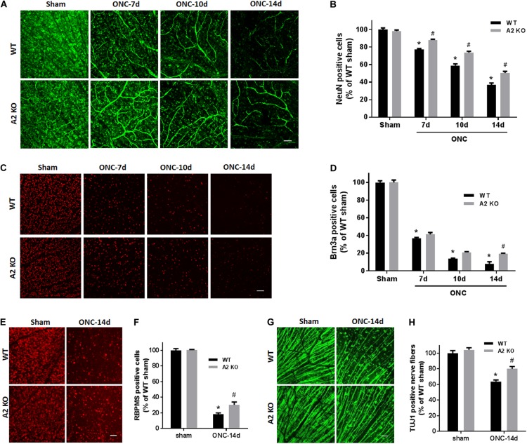
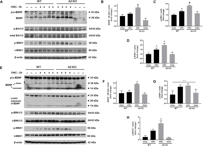
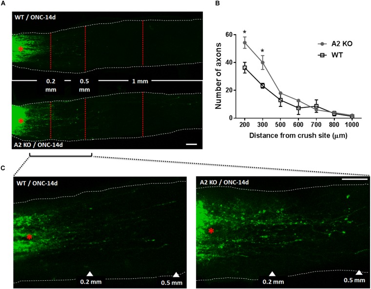
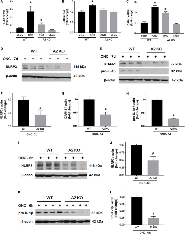
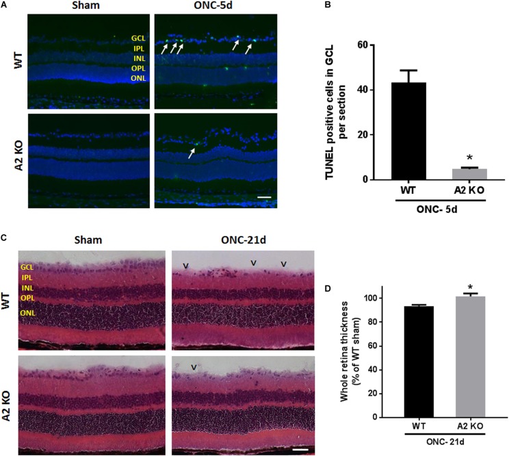
Our previous studies have implicated expression of the mitochondrial isoform of the arginase enzyme arginase 2 (A2) in neurovascular injury during ischemic retinopathies. The aim of this study was to characterize the specific involvement of A2 in retinal injury following optic nerve crush (ONC). To accomplish this, wild-type (WT) or A2 knockout (A2-/-) mice were subjected to ONC injury. The contralateral eye served as sham control. Quantitative RT-PCR and western blot were used to evaluate mRNA and protein expression. Retinal ganglion cell (RGC) survival was assessed in retinal whole mounts. Axonal sprouting was determined by anterograde transport of Cholera Toxin B (CTB). These analyses showed increased A2 expression following ONC. Numbers of NeuN-positive neurons as well as Brn3a- and RBPMS-positive RGC were decreased in the WT retinas at 14 days after ONC as compared to the sham controls. This ONC-induced neuronal loss was diminished in the A2-/- retinas. Similarly, axonal degeneration was ameliorated by A2 deletion whereas axon sprouting was enhanced. Significant retinal thinning was also seen in WT retinas at 21 days after ONC, and this was blocked in A2-/- mice. Cell death studies showed an increase in TUNEL positive cells in the RGC layer at 5 days after ONC in the WT retinas, and this was attenuated by A2 deletion. ONC increased glial cell activation in WT retinas, and this was significantly reduced by A2 deletion. Western blotting showed a marked increase in the neurotrophin, brain derived neurotrophic factor (BDNF) and its downstream signaling in A2-/- retinas vs. WT after ONC. This was associated with increases in the axonal regeneration marker GAP-43 in A2-/- retinas. Furthermore, A2-/- retinas showed decreased NLRP3 inflammasome activation and lower interleukin (IL-) 1β/IL-18 levels as compared to WT retinas subjected to ONC. Collectively, our results show that deletion of A2 limits ONC-induced neurodegeneration and glial activation, and enhances axonal sprouting by a mechanism involving increases in BDNF and decreases in retinal inflammation. These data demonstrate that A2 plays an important role in ONC-induced retinal damage. Blockade of A2 activity may offer a therapeutic strategy for preventing vision loss induced by traumatic retinal injury.

Keywords: arginase 2; brain derived neurotrophic factor; neuroprotection; optic nerve crush; retina; retinal ganglion cells.


Figures

FIGURE 1
FIGURE 1
A2 is increased in retinas subjected to ONC injury. Schematic diagram (A) of the different time points examined after subjecting WT and A2-/- mice to ONC. Western blotting (B) of retinal lysates for A1 and A2 protein expression and quantification (C,D) showed increases in A2 in response to ONC at 6 h after injury. A2-/- retinas lacked the A2 protein band. A1 expression was similar in all groups. N = 3–4, p < 0.01 vs. WT sham, n.s. means no statistically significant difference.
FIGURE 2
FIGURE 2
A2 deletion ameliorates ONC-induced neuronal cell loss. Representative NeuN labeling (A) and quantification (B) of retinal neurons in the GCL of retinal flat mounts show a progressive decline in NeuN+ cells over 14 days after ONC. A2 deletion ameliorates the ONC-induced neuronal cell loss. Immunolabeling (C) and quantification (D) for the RGC marker, Brna3a, showed a similar trend with reduced neurodegeneration in the A2-/- retinas. N = 4–6 per group, p < 0.01 vs. WT sham, #p < 0.01 vs. A2-/- sham and respective WT ONC time point. Flat mount labeling for another RGC marker, RBPMS, at 14 days (E) and quantification (F) showed similar results with significant neuroprotection in A2-/- retinas. Flat-mount labeling of the retinal neurons nerve fibers using TUJ1 (G) and quantification (H) showed nerve fiber loss at 14 days after ONC. A2 deletion was associated with relative preservation of nerve fibers as compared to WT after ONC. N = 5–6, p < 0.01 vs. WT sham, #p < 0.01 vs. A2-/- sham and WT ONC, scale bar = 50 μm.
FIGURE 3
FIGURE 3
A2 deletion increases mature BDNF and its downstream signaling targets after ONC. Western blotting on retinal lysates and quantification at 3 h (A–D) and 2 days (E–H) after injury showed increased expression of mature BDNF (28 kD) and activation of its downstream signaling pathway (ERK/MSK) in the A2-/- ONC retinas as compared to WT. N = 3–4, p < 0.05 vs. WT sham, #p < 0.05 vs. the other three groups, n.s. means no statistically significant difference.
FIGURE 4
FIGURE 4
A2 deletion increases GAP-43 expression in nerve fibers after ONC. Flat mount immunolabeling (A) showed higher GAP-43 expression in nerve fibers of WT ONC vs. sham retinas at 7 days and A2 deletion further increased GAP-43 immunoreactivity. N = 4–5, scale bar = 50 μm. Western blotting on retina lysates (B) and quantification (C) showed increased GAP-43 expression in A2-/- retinas as compared to WT that was significant at 5 days but did not reach statistical significance at 7 days after ONC. N = 3, p < 0.05 vs. WT ONC, n.s. means no statistically significant difference.
FIGURE 5
FIGURE 5
A2 deletion enhances axonal sprouting after ONC. Anterograde labeling of RGC axons using CTB (A) showed a significant increase in the number of axons that traveled beyond the crush site toward the brain at 200 and 300 μm distance from crush site denoted by asterisk (B), N = 3 per group, p < 0.05. (C) Shows higher magnification images of WT and A2-/- optic nerves from crush site to 500 μm distance toward the brain. Scale bar = 100 μm.
FIGURE 6
FIGURE 6
A2 deletion reduces glial activation after ONC. Immunolabeling for Iba1 in retina cross sections (A) and flat-mounts (B) together with quantification (C) showed prominent activation of microglia/macrophage (numerous Iba1+ cells with ameboid morphology) in WT retinas at 7 days after ONC. This was ameliorated in the A2-/- retinas and the Iba1+ cells had a ramified morphology. N = 4. Labeling for GFAP on cross sections (D) and quantification (E) showed increased glial activation at 7 days after ONC which was reduced with A2 deletion. N = 5. p < 0.01 vs. WT sham, #p < 0.01 vs. WT ONC. Arrows denote upregulation of GFAP in the radial processes of activated Müller cells. Scale bar = 50 μm.
FIGURE 7
FIGURE 7
A2 deletion ameliorates ONC-induced retinal inflammation. Quantitative RT-PCR (A–C) showed upregulation of pro-inflammatory mediators (IL-1β, IL-18, and ICAM-1) at 7 days after ONC in the WT retinas. A2 deletion significantly blunted these increases. N = 4–6, p < 0.05 vs. WT sham, #p < 0.05 vs. WT ONC. Western blotting at 7 days after ONC (D,E) and quantification (F–H) showed a significant reduction in NLRP3, ICAM-1, and pro-IL-1β protein levels in A2-/- retinas as compared to WT. Western blotting on retina lysates (I,K) and quantification (J,L) showed downregulation of NLRP3 and pro-IL-1β in A2-/- retinas as compared to WT at 6 h after ONC as well. N = 3–4, #p < 0.05 vs. WT ONC.
FIGURE 8
FIGURE 8
A2 deletion ameliorates ONC-induced apoptosis and limits retinal thinning. TUNEL labeling (A) of retina cross sections and quantification (B) showed increased TUNEL positive cells in the GCL (arrows) at 5 days after ONC. A2-/- retinas showed a significant reduction in TUNEL positive cells. N = 4–6 per group, p < 0.01, scale bar = 50 μm. Hematoxylin and eosin (H&E) staining of retina cross sections (C) and quantification (D) at 21 days showed that A2 deletion preserved the total retina thickness to near sham levels. N = 6 per group, p < 0.01, scale bar = 50 μm. Arrowheads denote area of RGC loss.

References

    1. Caldwell R. W., Rodriguez P. C., Toque H. A., Narayanan S. P., Caldwell R. B. (2018). Arginase: a multifaceted enzyme important in health and disease. Physiol. Rev. 98 641–665. 10.1152/physrev.00037.2016 - DOI - PMC - PubMed
    1. Doster S. K., Lozano A. M., Aguayo A. J., Willard M. B. (1991). Expression of the growth-associated protein GAP-43 in adult rat retinal ganglion cells following axon injury. Neuron 6 635–647. 10.1016/0896-6273(91)90066-9 - DOI - PubMed
    1. Fouda A. Y., Xu Z., Shosha E., Lemtalsi T., Chen J., Toque H. A., et al. (2018). Arginase 1 promotes retinal neurovascular protection from ischemia through suppression of macrophage inflammatory responses. Cell Death Dis. 9:1001. 10.1038/s41419-018-1051-6 - DOI - PMC - PubMed
    1. Fournier A. E., Beer J., Arregui C. O., Essagian C., Aguayo A. J., Mckerracher L. (1997). Brain-derived neurotrophic factor modulates GAP-43 but not T alpha1 expression in injured retinal ganglion cells of adult rats. J. Neurosci. Res. 47 561–572. 10.1002/(SICI)1097-4547(19970315)47:6<561::AID-JNR1>3.0.CO;2-B - DOI - PubMed
    1. Gupta S. K., Mishra R., Kusum S., Spedding M., Meiri K. F., Gressens P., et al. (2009). GAP-43 is essential for the neurotrophic effects of BDNF and positive AMPA receptor modulator S18986. Cell Death. Differ. 16 624–637. 10.1038/cdd.2008.188 - DOI - PubMed