. 2018 Dec 18:12:978.
doi: 10.3389/fnins.2018.00978. eCollection 2018.

Spatiotemporal Structure and Dynamics of Spontaneous Oscillatory Synchrony in the Vagal Complex

Affiliations

Spatiotemporal Structure and Dynamics of Spontaneous Oscillatory Synchrony in the Vagal Complex

Yoshinori Kawai. Front Neurosci. .

Abstract

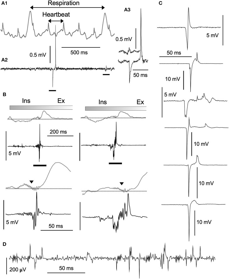
Fundamental structure and dynamics of spontaneous neuronal activities without apparent peripheral inputs were analyzed in the vagal complex (VC), whose activities had been generally thought to be produced almost passively to peripheral cues. The analysis included the caudal nucleus of the tractus solitarius-a main gateway for viscerosensory peripheral afferents and involved dynamically and critically in cardiorespiratory brainstem networks. In the present study, a possibility of self-organized brain activity was addressed in the VC. While VC neurons exhibited sparse firing in anesthetized rats and in in vitro preparations, we identified peculiar features of the emergent electrical population activity: (1) Spontaneous neuronal activity, in most cases, comprised both respiration and cardiac cycle components. (2) Population potentials of polyphasic high amplitudes reaching several millivolts emerged in synchrony with the inspiratory phase of respiratory cycles and exhibited several other characteristic temporal dynamics. (3) The spatiotemporal dynamics of local field potentials (LFPs), recorded simultaneously over multiple sites, were characterized by a stochastic emergence of high-amplitude synchrony. By adjusting amplitude and frequency (phase) over both space and time, the traveling synchrony exhibited varied degrees of coherence and power with a fluctuating balance between mutual oscillators of respiratory and cardiac frequency ranges. Full-fledged large-scale oscillatory synchrony over a wide region of the VC emerged after achieving a maximal stable balance between the two oscillators. Distinct somatic (respiratory; ~1 Hz) and visceral (autonomic; ~5 Hz) oscillators seemed to exist and communicate co-operatively in the brainstem network. Fluctuating oscillatory coupling may reflect varied degrees of synchrony influenced by the varied amplitude and frequency of neuronal activity in the VC. Intranuclear micro-, intrabulbar meso-, and wide-ranging macro-circuits involving the VC are likely to form nested networks and strategically interact to maintain a malleable whole-body homeostasis. These two brainstem oscillators could orchestrate neuronal activities of the VC, and other neuronal groups, through a phase-phase coupling mechanism to perform specific physiological functions.

Keywords: brain wave; electrical activity; emergence; oscillator; self-organization; viscerosensory.


Figures

Figure 1
Figure 1
In vivo (A) and in vitro (B) preparations for recordings of spontaneous neuronal activities from the vagal complex (VC). (A) The VC consists of the dorsal motor nucleus of the vagus (dmnX, layer III) and the nucleus of tractus solitarius (NTS, gray-shaded) that can be divided into dorsal and ventral (I and II) layers according to function and cytoarchitecture (Negishi and Kawai, 2011). A silicon electrode of 16 metal probes spanned the whole depth of the VC (a vertical scale in μm). (A1) is magnified from a frame in (A2). (B1) An upright ponto-medulla block preparation of newborn rats. (B2) Cerebellum was detached from this preparation. Amb, ambiguus nucleus; ap, area postrema; cc, central canal; cp, cerebellar peduncle (cut); Gr, gracilis nucleus; IC, inferior colliculus; nXII (12), hypoglossal nucleus; py, pyramidal tract; Vsp, spinal nucleus of the trigeminal nerves.
Figure 2
Figure 2
Structure of high-amplitude potentials. Neuronal high-amplitude potentials recorded extracellularly in vivo using a standard glass electrode in the vagal complex. (A1) Cardiorespiratory activities recorded simultaneously with a piezoelectric transducer (PZT) attached to a thorax (gray wave in an upper row). Cardiorespiratory cycles (“Respiration” and “Heartbeat” cycles indicated by double-headed arrows) were confirmed by a visual inspection of thorax movement. A simultaneously recorded neuronal activity (A2, lower row) contains a high-amplitude potential and a typical low-amplitude wave (A3, bars in A2). (B) Polyphasic high-amplitude potentials synchronized with the inspiratory (Ins) phase of each respiratory cycle. The polyphasic potential activity is reflected as synchronized jitter in cardiorespiratory PZT traces (triangles in lower rows expanded from bars in upper rows). Varied shapes of high-amplitude potentials ranging several millivolts in amplitude, as depicted in (B) (synchronized with respiratory cycles) and (C) (asynchronous with cardiorespiratory cycles), look like enlarged copies of typical low-amplitude potentials ranging hundred microvolts (D). Burst-like polyphasic activities are associated with both types of potentials. Ex, expiratory phase of a respiratory cycle.
Figure 3
Figure 3
Neuronal activities synchronized with cardiorespiratory cycles, recorded in vivo using a glass electrode. (A1) Episodic emergence of trains of high-amplitude potentials are synchronized with the respiratory cycle. PZT traces (A1–3; shown in gray) exhibit a respiratory cycle of ~1.1 Hz and a heartbeat cycle of ~5.6 Hz. Trains of high-amplitude potentials synchronized with respiratory cycles are marked with solid dots. Three episodes of a 3–4 s duration before, during, and after the high-amplitude potential trains, corresponding to horizontal bars in (A1) are shown in (A2) with simultaneously-recorded PZT traces. High-amplitude potential traces in the middle are truncated. Note the polyphasic high-amplitude potentials (solid triangles) synchronized with the inspiration phase (gray-shaded). The last episode contains low-amplitude and longer-duration signals during the inspiration phase (gray-shaded) in addition to short-duration spikes. (A3) PZT-trough-triggered high-amplitude potentials of 20 successive respiratory cycles. Multiple potentials appear during the inspiration phase (gray-shaded). (A4) and (A5) Power-spectrum analyses (Power) and a correlogram (Corr.) of neuronal and cardiorespiratory (PZT: gray in A4) activities are shown. Respiratory peaks of ~1 Hz are marked with solid circles. (B1) Low-amplitude spikes synchronized with heartbeat cycles. A simultaneous in vivo recording of neuronal and cardiorespiratory (in gray) activities indicate synchrony of short-duration spikes of ~100 μV amplitude with heartbeat cycles of ~5.9 Hz (solid circles). (B2) and (B3), Power-spectrum analyses (Power) and a correlogram (Corr.) of neuronal and cardiorespiratory (PZT: gray in B2) activities. Heartbeat and respiratory peaks are marked with solid and open circles, respectively. NTS, the nucleus tractus solitarius.
Figure 4
Figure 4
Spontaneous neuronal activities of the ventral vagal complex (vVC) synchronized with presumed respiratory rhythms recorded in an in vitro ponto-medullary block. (A) Simultaneous recordings of a vVC neuron and hypoglossal nerve rootlets (nXII; in gray) reveal synchronous activities (solid circles) between them. Expanded traces (A1 lower and A2) show concurrent single (vVC) and multiple (nXII) spikes. (B) Power spectrum analyses (Power) of vVC and nXII (in gray) activities over slow (B1) and slower (B2) frequency ranges. A vertical axis in (B2) is arbitrarily set for a comparison of peaks of the two sets of power. Synchronous peaks of power spectra are marked by solid circles (B1,2). (C) A correlogram (Corr.) of vVC and nXII activities.
Figure 5
Figure 5
Temporal dynamics of vagal complex (VC) neuronal activities synchronized with cardiorespiratory rhythms. (A) A piezoelectric pulse transducer (PZT) recorded cardiorespiratory rhythmic wave and the time-resolved power spectrum by the continuous wavelet transform (CWT) analysis (A1). Respiratory (1.65 Hz, black arrow) and cardiac (6.97 Hz, red arrow) rhythms are shown in the CWT time-resolved power spectrum. (A2) An FFT power spectrum of PZT consists of fundamental respiratory (1.65 Hz, black arrow) and cardiac (6.97 Hz, red arrow) frequency peaks and several respiratory harmonic peaks. (B) Temporal phase transition of polyphasic high-amplitude potentials occurring in synchrony with every cardiac cycle (B1), every respiratory cycle (B2), every second respiratory cycle (B3, open and solid dots), every third respiratory cycle (B4, open and solid dots), and every fourth respiratory cycle (B5, open and solid dots). (C) Ten-s-duration power spectra (C1–5) corresponding to (B1–5) recordings, respectively. Arrows (black and red) indicate respiratory and cardiac frequency components corresponding to those in PZT (A2).
Figure 6
Figure 6
A simultaneous multiple recording of local field potentials (LFPs). (A) A two-row silicon probe consisting of 16 electrodes each separated by 50 μm in length and 400 μm in depth, in the vagal complex (VC). (B) An example of LFPs of 100 s duration from eight vertically arranged sites (gray in A). (C) An expanded 10 s-duration-LFPs from a period of 70–80 s in (B) (gray rectangle). (D) CWT time-resolved power spectrum results corresponding to this 10-s period of simultaneous recordings. Note that lower-amplitude LFPs in the dorsal VC have more intense signals of respiratory frequency range (~1 Hz) and higher-amplitude signals in the ventral cardiac range (~5 Hz). The spatial wave structure shaped by differential frequency ranges fluctuates temporally over 100 s and higher-amplitude waves are noted in deeper layers (II and III) (B). CWT, continuous wavelet transform.
Figure 7
Figure 7
Spatiotemporal dynamics of wave correlation (Corr.) and coherence of multiple local field potentials (LFPs). Wave Corr. and coherence of a 10 s duration between LFPs recorded 30 s apart from neighboring pairs of electrodes (gray shades in A1 and B1) across the depth of the vagal complex. Note a tendency of temporally-upward (gray arrow in A1) or -downward (gray arrow in B1) increases in Corr. (3 colored pairs in A2,B2) and an apparent higher coherence during 1–5 Hz frequency range (3 colored pairs in A3,B3).
Figure 8
Figure 8
Continuous wavelet transform (CWT) time-resolved power spectrum profiles predict signal amplitude and frequency. (A) Dynamics of vagal complex (VC) local field potentials (LFPs) obtained offline by digitally-low pass filtering (LP) in synchrony with piezoelectric pulse transducer (PZT) signals. (A1) LP signals (gray in the lowest trace) obtained from original high-amplitude potentials in synchrony with every inspiration phase (middle traces) and PZT signals (gray in the upper trace) produce similar CWT structures. (A2) LP and PZT signals give different CWT profiles. Compared to PZT, the LP CWT profile has intense signals for larger frequencies (> ~5 Hz) and very low signals for ~1 Hz frequencies. Note that amplitudes of LP signals are far larger than those shown in (A1), and the VC high-amplitude signals synchronize with every second respiratory component of the PZT signals. Time-resolved coherence spectra (wavelet coherence; Wcoh) between PZT and LP signals show different profiles with either ~1 Hz or ~1/~3 Hz dense bands (white arrows). (B) and (C), CWT profiles (upper in B1,C1) and the corresponding VC LFPs (lower in B1,C1) recorded with a multiple silicon electrode. (B1) Two episodes of recordings from the same site temporally 5 s apart. (B2) Power spectrum measurements of each episode over a period of ~5 s. (C1) Two simultaneous recordings from two sites spatially 300 μm apart. (C2) Correlograms (Corr.) of each episode between adjacent LFPs (50 μm apart). (D1) and (D2) Linear correlation between Corr. of two simultaneously-recorded LFPs according to the mean signal amplitudes (D1) and the distances between recording sites (D2).
Figure 9
Figure 9
Large-scale spontaneous oscillatory synchrony. (A1) Three successive episodes (green, orange, red) of adjacent LFP pairs (50 μm apart) recorded by a silicon multiple electrode. (A2) In the third episode, all the LFPs of similar trajectories emerged, ensued, and then disappeared. (A3) Superimposition of the episodes. (B) Time-resolved power spectra (CWT) of the episodes. (C) Power spectra of the episodes (each color same with A). Note two power peaks (solid dots; ~1 and ~5 Hz) in the large-scale synchrony (red). (D) Coherence spectra of the episodes. (E1) and (E2), Superimposition of an autocorrelogram and a correlogram between adjacent paired waves of the three episodes. Note that oscillatory frequencies differ among the episodes and a salient mixture of two oscillation modes (~1 and ~5 Hz) in the last episode (E2). (F) Time-resolved coherence spectra (wavelet coherence; Wcoh) between PZT and LFP signals according to an amplitude of LFPs. As LFP amplitudes develop (left to right), dense bands of high coherence occupy a wider range of frequency. PZT, piezoelectric pulse transducer; LFP, local field potential.

Similar articles

Cited by

References

    1. Bianchi A. L., Denavit-Saubie M., Champagnat J. (1995). Central control of breathing in mammals: neuronal circuitry, membrane properties, and neurotransmitters. Physiol. Rev. 75, 1–45. 10.1152/physrev.1995.75.1.1 - DOI - PubMed
    1. Blanche T. J., Spacek M. A., Hetke J. F., Swindale N. V. (2005). Polytrodes: high-density silicon electrode arrays for large-scale multiunit recording. J. Neurophysiol. 93, 2987–3000. 10.1152/jn.01023.2004 - DOI - PubMed
    1. Boczek-Funcke A., Dembowsky K., Habler H. J., Janig W., Michaelis M. (1992). Respiratory-related activity patterns in preganglionic neurones projecting into the cat cervical sympathetic trunk. J. Physiol. 457, 277–296. 10.1113/jphysiol.1992.sp019378 - DOI - PMC - PubMed
    1. Buzsaki G. (2006). Rhythms of the Brain. New York, NY: Oxford University Press, Inc; 10.1093/acprof:oso/9780195301069.001.0001 - DOI
    1. Canolty R. T., Knight R. T. (2010). The functional role of cross-frequency coupling. Trends Cogn. Sci. 14, 506–515. 10.1016/j.tics.2010.09.001 - DOI - PMC - PubMed

LinkOut - more resources