Review
. 2018 Dec 17:11:468.
doi: 10.3389/fnmol.2018.00468. eCollection 2018.

The Complex Conformational Dynamics of Neuronal Calcium Sensor-1: A Single Molecule Perspective

Affiliations
Review

The Complex Conformational Dynamics of Neuronal Calcium Sensor-1: A Single Molecule Perspective

Dhawal Choudhary et al. Front Mol Neurosci. .

Abstract

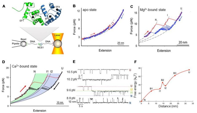
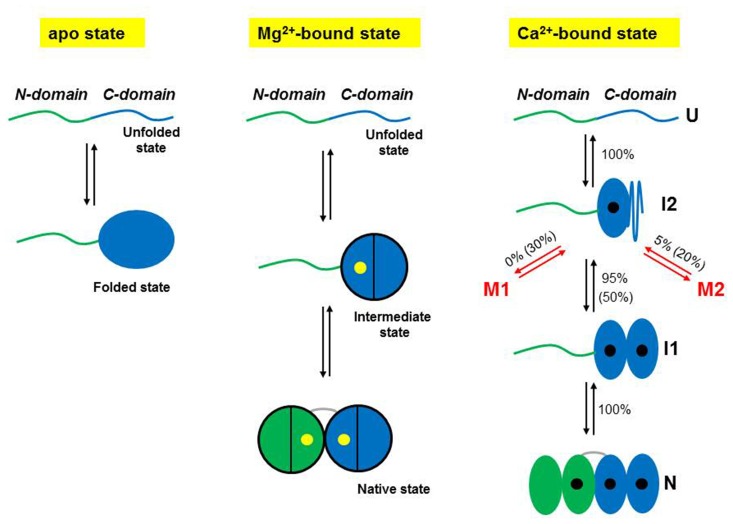
The human neuronal calcium sensor-1 (NCS-1) is a multispecific two-domain EF-hand protein expressed predominantly in neurons and is a member of the NCS protein family. Structure-function relationships of NCS-1 have been extensively studied showing that conformational dynamics linked to diverse ion-binding is important to its function. NCS-1 transduces Ca2+ changes in neurons and is linked to a wide range of neuronal functions such as regulation of neurotransmitter release, voltage-gated Ca2+ channels and neuronal outgrowth. Defective NCS-1 can be deleterious to cells and has been linked to serious neuronal disorders like autism. Here, we review recent studies describing at the single molecule level the structural and mechanistic details of the folding and misfolding processes of the non-myristoylated NCS-1. By manipulating one molecule at a time with optical tweezers, the conformational equilibria of the Ca2+-bound, Mg2+-bound and apo states of NCS-1 were investigated revealing a complex folding mechanism underlain by a rugged and multidimensional energy landscape. The molecular rearrangements that NCS-1 undergoes to transit from one conformation to another and the energetics of these reactions are tightly regulated by the binding of divalent ions (Ca2+ and Mg2+) to its EF-hands. At pathologically high Ca2+ concentrations the protein sometimes follows non-productive misfolding pathways leading to kinetically trapped and potentially harmful misfolded conformations. We discuss the significance of these misfolding events as well as the role of inter-domain interactions in shaping the energy landscape and ultimately the biological function of NCS-1. The conformational equilibria of NCS-1 are also compared to those of calmodulin (CaM) and differences and similarities in the behavior of these proteins are rationalized in terms of structural properties.

Keywords: NCS-1; calcium binding; optical tweezers; protein folding and misfolding; single molecule studies.


Figures

Figure 1
Figure 1
Mechanical manipulation of a single non-myristoylated neuronal calcium sensor-1 (NCS-1) molecule. (A) Schematic representation of the optical tweezers setup. A NCS-1 molecule is tethered to two polystyrene beads by means of molecular handles (~500 bp DNA molecules) that function as spacers to avoid unspecific interaction between the tethering surfaces (Cecconi et al., 2005). One bead is held in an optical trap, while the other is held at the end of a pipette by suction. During the experiment the protein is stretched and relaxed by moving the pipette relative to the optical trap, and the applied force and the molecular extension are measured as described in Smith et al. (2003). The inset shows the NMR structure of NCS-1 (PDB code 2LCP), where the C- and N-domains are shown in blue and green, respectively. (B) Force vs. extension cycle obtained by stretching (red trace) and relaxing (blue trace) NCS-1 in the absence of divalent ions. (C) Force vs. extension cycle obtained by stretching (red trace) and relaxing (blue trace) NCS-1 in the presence of 10 mM Mg2+. (D) Mechanical manipulation of the Ca2+-bound state of NCS-1. During stretching (red trace) the N-domain unfolds at ~13 pN and the C-domain at ~16 pN. During relaxation (black trace), U folds into N through a four-state process (U > I2 > I1 > N) coordinated by calcium binding. (E) Extension vs. time traces acquired at different constant forces showing the Ca2+-bound state of NCS-1 fluctuating at equilibrium between N, I1, I2 and U. The population probability of the different states can be modulated by force. Analysis of these experimental data with the Hidden Markov Model allows for the characterization of the energy landscape of the protein in terms of activation energy barriers, separating the different molecular states, and positions of the transition states along the reaction coordinate (Rabiner, ; Chodera et al., 2011). (F) Energy landscape of the Ca2+-bound state of NCS-1 at zero force. The transition states of the different folding reactions are indicated with the letter B. The activation barriers separating the different molecular states were calculated using a pre-exponential factor of 1.2 × 10⁻⁴ Hz (Gebhardt et al., 2010). Panels adapted from Heidarsson et al. (2013b) and Naqvi et al. (2015).
Figure 2
Figure 2
Schematic representations of the folding mechanisms of non-myristoylated NCS-1 in different ionic conditions. In the absence of divalent ions, the C-domain fluctuates between a folded and an unfolded state, while the N-domain remains unstructured or loosely folded. In the presence of Mg2+ (yellow dots), the divalent ions binds first to EF3, triggering the folding of the C-domain, and then to EF2 making the NCS-1 transit into its native state. Only two of the three active EF-hands can bind Mg2+. Under activating Ca2+ concentrations, NCS-1 folds into its native state through a four-state process involving the population of the intermediate states I1 and I2. First NCS-1 transits into I2 and then into I1 as Ca2+ (black dots) binds to EF3 and EF4, respectively. Finally Ca2+ binds to EF2 and the protein reaches N. At high calcium concentration (10 mM), only one molecule out of two follows this native folding pathway. Fifty percentage of the molecules folds into I2 and then takes non-productive pathways leading to misfolded conformations (M1 and M2). In contrast, at physiological calcium concentrations (0.5 μM), only 5% of the molecules misfold. Panels adapted from Heidarsson et al. (2014) and Naqvi et al. (2015).

References

    1. Alemany A., Rey-Serra B., Frutos S., Cecconi C., Ritort F. (2016). Mechanical folding and unfolding of protein barnase at the single-molecule level. Biophys. J. 110, 63–74. 10.1016/j.bpj.2015.11.015 - DOI - PMC - PubMed
    1. Ames J. B., Hendricks K. B., Strahl T., Huttner I. G., Hamasaki N., Thorner J. (2000). Structure and calcium-binding properties of Frq1, a novel calcium sensor in the yeast Saccharomyces cerevisiae. Biochemistry 39, 12149–12161. 10.1021/bi0012890 - DOI - PubMed
    1. Aravind P., Chandra K., Reddy P. P., Jeromin A., Chary K., Sharma Y. (2008). Regulatory and structural EF-hand motifs of neuronal calcium sensor-1: Mg2+ modulates Ca2+ binding, Ca2+-induced conformational changes, and equilibrium unfolding transitions. J. Mol. Biol. 376, 1100–1115. 10.1016/j.jmb.2007.12.033 - DOI - PubMed
    1. Avdoshenko S. M., Makarov D. E. (2016). Reaction coordinates and pathways of mechanochemical transformations. J. Phys. Chem. B 120, 1537–1545. 10.1021/acs.jpcb.5b07613 - DOI - PubMed
    1. Bai J., He F., Novikova S. I., Undie A. S., Dracheva S., Haroutunian V., et al. . (2004). Abnormalities in the dopamine system in schizophrenia may lie in altered levels of dopamine receptor-interacting proteins. Biol. Psychiatry 56, 427–440. 10.1016/j.biopsych.2004.06.022 - DOI - PubMed