. 2018 Dec 11:9:1383.
doi: 10.3389/fphar.2018.01383. eCollection 2018.

Neuroprotective Effect of Quercetin Against the Detrimental Effects of LPS in the Adult Mouse Brain

Affiliations

Neuroprotective Effect of Quercetin Against the Detrimental Effects of LPS in the Adult Mouse Brain

Amjad Khan et al. Front Pharmacol. .

Abstract

Chronic neuroinflammation is responsible for multiple neurodegenerative diseases, such as Alzheimer's disease, Parkinson's disease, and Huntington's disease. Lipopolysaccharide (LPS) is an essential component of the gram-negative bacterial cell wall and acts as a potent stimulator of neuroinflammation that mediates neurodegeneration. Quercetin is a natural flavonoid that is abundantly found in fruits and vegetables and has been shown to possess multiple forms of desirable biological activity including anti-inflammatory and antioxidant properties. This study aimed to evaluate the neuroprotective effect of quercetin against the detrimental effects of LPS, such as neuroinflammation-mediated neurodegeneration and synaptic/memory dysfunction, in adult mice. LPS [0.25 mg/kg/day, intraperitoneally (I.P.) injections for 1 week]-induced glial activation causes the secretion of cytokines/chemokines and other inflammatory mediators, which further activate the mitochondrial apoptotic pathway and neuronal degeneration. Compared to LPS alone, quercetin (30 mg/kg/day, I.P.) for 2 weeks (1 week prior to the LPS and 1 week cotreated with LPS) significantly reduced activated gliosis and various inflammatory markers and prevented neuroinflammation in the cortex and hippocampus of adult mice. Furthermore, quercetin rescued the mitochondrial apoptotic pathway and neuronal degeneration by regulating Bax/Bcl2, and decreasing activated cytochrome c, caspase-3 activity and cleaving PARP-1 in the cortical and hippocampal regions of the mouse brain. The quercetin treatment significantly reversed the LPS-induced synaptic loss in the cortex and hippocampus of the adult mouse brain and improved the memory performance of the LPS-treated mice. In summary, our results demonstrate that natural flavonoids such as quercetin can be beneficial against LPS-induced neurotoxicity in adult mice.

Keywords: activated gliosis; lipopolysaccharide; memory performance; natural flavonoids; neuroinflammation; neurotoxicity; quercetin.


Figures

FIGURE 1
FIGURE 1
Schematic of the experimental schedule. (1) Control paradigm mice (Cont) treated with saline as vehicle for 2 weeks. (2) Mice treated with vehicle for 1 week and LPS 0.25 mg/kg/day for 1 week. (3) Mice treated with LPS 0.25 mg/kg/day for 1 week and quercetin 30 mg/kg/day for 2 weeks (1 week before the LPS and 1 week cotreated with LPS). After behavioral analyses on the 14th day, the adult mice were euthanized and further subjected to western blotting and morphological analyses.
FIGURE 2
FIGURE 2
Quercetin improved the memory function of the LPS-treated mice. For the behavioral analyses, the MWM and Y-maze tests were used to investigate and evaluate the memory functions of the control, LPS and LPS+quercetin group mice. (A) Average escape latency time for experimental mice to reach the hidden platform from 1 to 4 days. (B) The average number of crossings at the hidden platform during the probe test of the MWM test. (C) Time spent in the platform quadrant, where the hidden platform was placed during the trial session. (D) Spontaneous alteration behavior % of the mice during the Y-maze test. Histograms indicate the means ± SEM for the mice (n = 15/group). Significantly different from the control; # significantly different from LPS-treated group. Significance: P < 0.05.
FIGURE 3
FIGURE 3
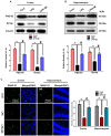
Quercetin improved the pre- and postsynaptic markers in the LPS-treated mice. (A,B) Western blotting of the proteins Synap and PSD95; their differences in the cortex and hippocampus of mouse brains are represented by a histogram. β-Actin was used as a loading control. The quantified density values are shown in arbitrary units (A.U.) as the means ± SEM for the respective shown protein (8 mice/group). (C) Representative immunofluorescence images and the quantified histogram of SNAP-23 in the cortex and hippocampus of adult mice (5 mice/group). Magnified 10×. Scale bar = 50 μm. The expressed data are relative to the control. Significantly different from the control; # significantly different from LPS-treated group. Significance: P < 0.05.
FIGURE 4
FIGURE 4
Quercetin ameliorated LPS-induced activated gliosis in the cortex and hippocampus of adult mice. (A,B) Western blotting of the proteins GFAP and Iba-1; their differences in the cortex and hippocampus of mouse brains are represented by a histogram. β-Actin was used as a loading control. The quantified density values are shown in arbitrary units (A.U.) as the means ± SEM for the respective shown protein (8 mice/group). (C) Representative immunofluorescence images and the quantified histogram of activated GFAP in the cortex and hippocampus of adult mice (5 mice/group). Magnified 10×. Scale bar = 50 μm. The expressed data are relative to the control. Significantly different from the control; # significantly different from LPS-treated group. Significance: P < 0.05.
FIGURE 5
FIGURE 5
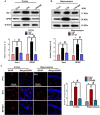
Quercetin halts the LPS-induced activated TLR4/NFKB pathway. (A,B) Western blotting of TLR4 and p-NFKB proteins; their differences in the cortex and hippocampus of mouse brains are represented by a histogram. β-Actin was used as a loading control. The quantified density values are shown in arbitrary units (A.U.) as the means ± SEM for the respective shown protein (8 mice/group). (C) Representative immunofluorescence images and the quantified histogram of p-NFKB in the cortex and hippocampus of adult mice (5 mice/group). Magnified 10×. Scale bar = 50 μm. The expressed data are relative to the control. Significantly different from the control; # significantly different from LPS-treated group. Significance: P < 0.05.
FIGURE 6
FIGURE 6
Quercetin attenuated LPS-induced neuroinflammation-associated markers. (A,B) Western blotting of TNF-α, COX-2 and NOS2 proteins; their differences in the cortex and hippocampus of mouse brains are represented by a histogram. β-Actin was used as a loading control. The quantified density values are shown in arbitrary units (A.U.) as the means ± SEM for the respective shown protein (8 mice/group). (C) Representative immunofluorescence images and the quantified histogram of IL-1β in the cortex and hippocampus of adult mice (5 mice/group). Magnified 10×. Scale bar = 50 μm. The expressed data are relative to the control. Significantly different from the control; # significantly different from LPS-treated group. Significance: P < 0.05.
FIGURE 7
FIGURE 7
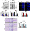
LPS-induced neuronal apoptotic pathway prevented by quercetin in the cortex and hippocampus. (A,B) Western blotting of Bcl-2, Bax, Cyto. c, Caspase-3 and PARP-1 proteins; their differences in the cortex and hippocampus of mouse brains are represented by a histogram. β-Actin was used as a loading control. The quantified density values are shown in arbitrary units (A.U.) as the means ± SEM for the respective shown protein (8 mice/group). (C) Representative immunofluorescence images and the quantified histogram of Caspase-3 in the cortex and hippocampus of adult mice (5 mice/group). Magnified 10×. Scale bar = 50 μm. (D) Representative immunohistochemical images (Nissl staining) and the quantified histogram of the survival neuron reactivity and integrated density in the cortex and hippocampus region of adult mice. The expressed data are relative to the control. Significantly different from the control; # significantly different from LPS-treated group. Significance: P < 0.05.

References

    1. Ahmad A., Ali T., Park H. Y., Badshah H., Rehman S. U., Kim M. O. (2016). Neuroprotective effect of fisetin against amyloid beta-induced cognitive/synaptic dysfunction, neuroinflammation and neurodegeneration in adult mice. Mol. Neurobiol. 54 2269–2285. 10.1007/s12035-016-9795-4 - DOI - PubMed
    1. Ali T., Badshah H., Kim T. H., Kim M. O. (2015a). Melatonin attenuates D-galactose-induced memory impairment, neuroinflammation and neurodegeneration via RAGE/NF-K B/JNK signaling pathway in aging mouse model. J. Pineal Res. 58 71–85. 10.1111/jpi.12194 - DOI - PubMed
    1. Ali T., Yoon G. H., Shah S. A., Lee H. Y., Kim M. O. (2015b). Osmotin attenuates amyloid beta-induced memory impairment, tau phosphorylation and neurodegeneration in the mouse hippocampus. Sci. Rep. 5:11708. 10.1038/srep11708 - DOI - PMC - PubMed
    1. Ali T., Kim T., Rehman S. U., Khan F. U., Khan M. Ikram M., et al. (2018). Natural dietary supplementaion of anthocyanins via PI3K/Akt/Nrf2/HO-1 pathways mitigates oxidative stress, neurodegeneration, and memory impairment in a mouse model of Alzheimer’s disease. Mol. Neurobiol. 55 6076–6093. 10.1007/s12035-017-0798-6 - DOI - PubMed
    1. Aravalli R. N., Peterson P. K., Lokensgard J. R. (2007). Toll-like receptors in defense and damage of the central nervous system. J. NeuroImmune Pharmacol. 2 297–312. 10.1007/s11481-007-9071-5 - DOI - PubMed