. 2018 Dec 12:9:3080.
doi: 10.3389/fmicb.2018.03080. eCollection 2018.

AU-Rich Long 3' Untranslated Region Regulates Gene Expression in Bacteria

Affiliations

AU-Rich Long 3' Untranslated Region Regulates Gene Expression in Bacteria

Ju-Ping Zhao et al. Front Microbiol. .

Abstract

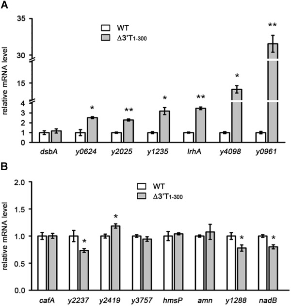
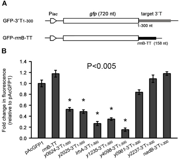
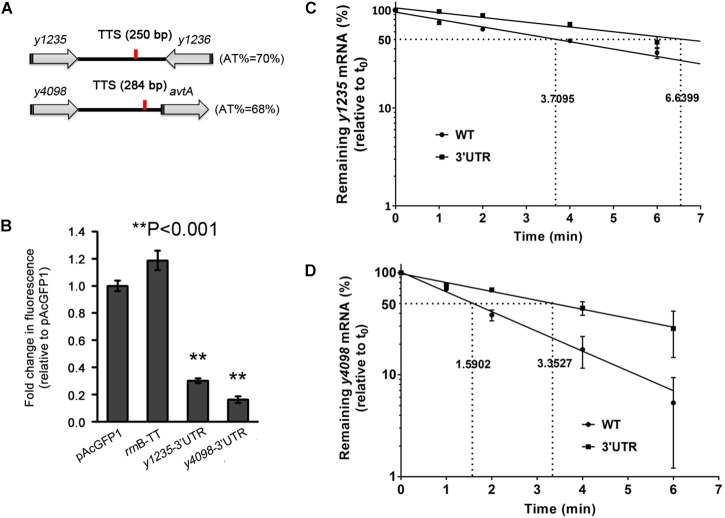
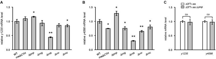
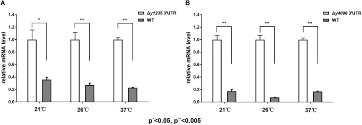
3' untranslated regions (3' UTRs) and particularly long 3' UTRs have been shown to act as a new class of post-transcriptional regulatory element. We previously reported that hmsT mRNA stability is negatively regulated by the 3' UTR of hmsT in Yersinia pestis. To investigate more general effects of 3' UTRs in Y. pestis, we selected 15 genes potentially possessing long 3' UTRs with different AU content and constructed their 3' UTR deletion mutants. Deletion of AU-rich 3' UTRs increased mRNA levels, whereas deletion of 3' UTRs with normal AU content resulted in slight or no changes in the mRNA level. In addition, we found that PNPase was important for 3' UTR-mediated mRNA decay when the transcriptional terminator was Rho-dependent. Finally, we showed that ribosomes promote mRNA stability when bound to a 3' UTR. Our findings suggest that functional 3' UTRs might be broadly distributed in bacteria and their novel regulatory mechanisms require further investigation.

Keywords: 3′ UTR; AU-rich region; Yersinia pestis; mRNA stability; post-transcriptional regulation.


Figures

FIGURE 1
FIGURE 1
Characterization of the general effects of 3′ UTRs on mRNA expression in Yersinia pestis. (A,B) qRT-PCR analysis of transcripts of genes containing AT-rich (A) or normal (B) 3′ IGRs in the wild type (WT) and nucleotides 1–300 of the 3′ terminus (3′ T1-300) mutant strains (Δ3′T1-300), in which the 3′ T1-300 is disrupted by insertion of a kanamycin (kan) cassette. The Y. pestis WT strain was used as a control, and the fold change in relative mRNA level for each strain was compared with that of the WT (set as 1). Student’s t-test was used to calculate p-values. p < 0.05, ∗∗p < 0.001. Means ± standard deviation (SD) from three independent experiments are shown.
FIGURE 2
FIGURE 2
The effects of 3′ UTRs on expression of a heterologous green fluorescent (GFP) reporter. (A) Schematic representation of GFP reporter with different 3′ UTRs and an rrnB transcriptional terminator. (B) Relative GFP fluorescence of E. coli cells expressing GFP with different 3′ UTRs (GFP-3′T1-300) and the rrnB terminator, and without an additional 3′ UTR (pAcGFP1). Relative fluorescence intensities were normalized against the cell density (OD600 value) of the bacterial culture, and the fold change in fluorescence was compared with that of the vector control (set as 1). Student’s t-tests were used to calculate p-values. p < 0.01. Means ± SD from three independent experiments are indicated.
FIGURE 3
FIGURE 3
The 3′ UTRs of y1235 and y4098 negatively regulate mRNA stability. (A) Schematic representation of y1235, y4098, and their transcriptional terminator (TTS, red line). (B) Relative GFP fluorescence of E. coli cells expressing GFP with the 3′ UTR of y1235 or y4098, or the rrnB terminator, or without an additional 3′ UTR (pAcGFP1). Relative fluorescence intensities were normalized against the OD600 value of the bacterial culture, and the fold change in fluorescence was compared with that of the vector control (set as 1). Student’s t-tests were used to calculate p-values. ∗∗p < 0.001. Means ± SD from three independent experiments are indicated. (C,D) The half-life of y1235 and y4098 mRNAs in WT and Δ3′ UTR mutant strains. Cells were grown at 26°C to an OD600 of 0.8, and rifampicin was added at time 0 to block RNA transcription. Samples were removed at time 0, 1 min, 2 min, 4 min, and 6 min. The percentage of residual mRNA at each time-point was measured by qRT-PCR and compared with that at time 0. Dashed lines indicate the time at which half of the detected mRNA remained. mRNA half-life was determined using linear regression analysis and is shown on the dashed lines. Error bars indicate SD of the mean.
FIGURE 4
FIGURE 4
Examination of the roles of Hfq in 3′ UTR-mediated decay of y1235 and y4098 mRNA. Relative amounts of y1235 (A) and y4098 (B) mRNA produced by the WT, Δ3′ UTR, Δhfq, and Δ3′ UTR Δhfq strains. Student’s t-test was used to calculate p-values. p < 0.01. Means ± standard deviation (SD) from three independent experiments are shown.
FIGURE 5
FIGURE 5
The effects of the major RNases on 3′ UTR-mediated mRNA turnover. The major RNases coding genes, CTH of rne [the carboxyl terminal half (CTH) region of RNase E], pnp (PNPase), rng (RNase G), rnb (RNase II), rnr (RNase R), and rnc (RNase III) were deleted, respectively. (A,B) Relative y1235 (A) and y4098 (B) mRNA levels in Y. pestis RNases mutants. (C) Relative y1235 and y4098 mRNA levels in Y. pestis Δ3′ UTR (white bars) and PNPase-Δ3′ UTR (gray bars) isogenic strains. Student’s t-test was used to calculate p-values. p < 0.05, ∗∗p < 0.001. Means ± SD from three independent experiments are indicated.
FIGURE 6
FIGURE 6
The effects of temperature on 3′ UTR-mediated mRNA turnover. qRT-PCR analysis of relative y1235 (A) and y4098 (B) mRNA levels in the WT and Δ3′ UTR mutant strain at 21°C, 26°C, and 37°C. Student’s t-test was used to calculate p-values. p < 0.01, ∗∗p < 0.005. Means ± SD from three independent experiments are indicated.
FIGURE 7
FIGURE 7
The effects of ribosome binding to 3′ UTR on mRNA decay. (A) Schematic representation of mutations introduced in 3′ UTRs of hmsT, y1235, and y4098. (B) qRT-PCR analysis of relative hmsT, y1235, and y4098 mRNA levels in Y. pestis WT strain, in the strain with a 3′ UTR containing a ribosome binding site (RBS), and in the strain containing the 3′ UTR with a sequence not recognized by the ribosome (RBSx). Student’s t-test was used to calculate p-values. p < 0.05. Means ± SD from three independent experiments are presented. (C) hmsT mRNA half-lives in the WT and 3′ UTR with RBS strains. Cells were grown at 26°C to an OD600 of ∼0.8, and rifampicin was added at time 0 to block RNA transcription. Samples were removed at time 0, 0.5 min, 1 min, 2 min, and 4 min. The percentage of mRNA remaining at each time-point was measured by qRT-PCR and compared with that at time 0. Dashed lines indicate the time at which half of the detected mRNA remained. The half-life of mRNAs was determined by linear regression analysis and is shown on the dashed lines. Error bars indicate the SD of the mean.

Similar articles

Cited by

References

    1. Agaisse H., Lereclus D. (1996). STAB-SD: a shine-dalgarno sequence in the 5’ untranslated region is a determinant of mRNA stability. Mol. Microbiol. 20 633–643. 10.1046/j.1365-2958.1996.5401046.x - DOI - PubMed
    1. Balaban N., Novick R. P. (1995). Translation of RNAIII, the Staphylococcus aureus agr regulatory RNA molecule, can be activated by a 3’-end deletion. FEMS Microbiol. Lett. 133 155–161. 10.1016/0378-1097(95)00356-A - DOI - PubMed
    1. Bechhofer D. H. (2009). Messenger RNA decay and maturation in Bacillus subtilis. Prog. Mol. Biol. Transl. Sci. 85 231–273. 10.1016/S0079-6603(08)00806-4 - DOI - PubMed
    1. Bellows L. E., Koestler B. J., Karaba S. M., Waters C. M., Lathem W. W. (2012). Hfq-dependent, co-ordinate control of cyclic diguanylate synthesis and catabolism in the plague pathogen Yersinia pestis. Mol. Microbiol. 86 661–674. 10.1111/mmi.12011 - DOI - PMC - PubMed
    1. Bouvier M., Carpousis A. J. (2011). A tale of two mRNA degradation pathways mediated by RNase E. Mol. Microbiol. 82 1305–1310. 10.1111/j.1365-2958.2011.07894.x - DOI - PubMed

LinkOut - more resources