. 2018 Dec 13:9:3104.
doi: 10.3389/fmicb.2018.03104. eCollection 2018.

Relevance of Bacteriophage 933W in the Development of Hemolytic Uremic Syndrome (HUS)

Affiliations

Relevance of Bacteriophage 933W in the Development of Hemolytic Uremic Syndrome (HUS)

Manuel E Del Cogliano et al. Front Microbiol. .

Abstract

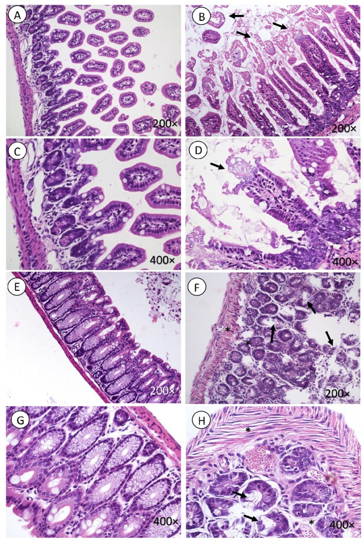
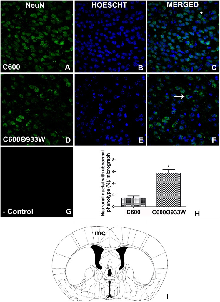
Hemolytic uremic syndrome (HUS), principally caused by shiga toxins (Stxs), is associated with Shiga toxin-producing Escherichia coli (STEC) infections. We previously reported Stx2 expression by host cells in vitro and in vivo. As the genes encoding the two Stx subunits are located in bacteriophage genomes, the aim of the current study was to evaluate the role of bacteriophage induction in HUS development in absence of an E. coli O157:H7 genomic background. Mice were inoculated with a non-pathogenic E. coli strain carrying the lysogenic bacteriophage 933W (C600Φ933W), and bacteriophage excision was induced by an antibiotic. The mice died 72 h after inoculation, having developed pathogenic damage typical of STEC infection. As well as renal and intestinal damage, markers of central nervous system (CNS) injury were observed, including aberrant immunolocalization of neuronal nuclei (NeuN) and increased expression of glial fibrillary acidic protein (GFAP). These results show that bacteriophage 933W without an E. coli O157:H7 background is capable of inducing the pathogenic damage associated with STEC infection. In addition, a novel mouse model was developed to evaluate therapeutic approaches focused on the bacteriophage as a new target.

Keywords: Shiga toxigenic E. coli (STEC); Shiga toxin (Stx); animal model; bacterio(phages); hemolytic uremic syndrome.


Figures

Figure 1
Figure 1
(A) Growth curves of E. coli C600 and E. coli C600Φ933W. Both strains present a similar growth curve with a non-significant difference between the viability counts. (B) Survival curve. Survival rates of mice infected with E. coli C600Φ933W are shown. Mice infected with E. coli C600 were used as controls. (C) Stx-induced renal damage. Plasmatic urea levels at 72 h post-infection were measured as a parameter of renal damage. Each bar represents the mean ± SEM of 4–6 mice/group. **P = 0.0095.
Figure 2
Figure 2
Kidney histology. Mesangial proliferation is observed in in mice infected with E. coli C600Φ933W (black arrow in A,C), as well as necrosis and detachment of tubular cells (black arrowhead in A,C), and tubular dilation (black asterisk in A,C). No effect was observed in mice infected eith the control strain E. coli C600 (B,D). Periodic acid shift (PAS). Magnification: 200 ×.
Figure 3
Figure 3
Intestine histology. Necrosis is observed in intestinal microvilli (black arrow, B,D) of the small intestine and in the crypt of the large intestine (black arrow in F,H). There is also marked edema surrounding the crypts of the mucosa and muscle fibers (black asterisk in F,H). (A,C,E,G) corresponding to the control group, did not present lesions. H&E. Magnification: (A,B and E,F 200 ×, C,D and G,H) 400 ×.
Figure 4
Figure 4
Detection of Stx2 in brains of mice infected with E. coli C600Φ933W. (A–C) A representative brain section from control mouse infected with E. coli C600. (D–F) Brain section from mouse infected with E. coli C600Φ933W. (G–I) Brain section from a positive control mouse after 4 days of treatment with 1 μg of Stx2. (J) Negative control. (K) Number of Stx2-immunopositive cells in in the mouse motor cortex (mc) per micrograph. Stx2 immunopositive cells are indicated by arrows in (F,I). (L) The area observed is located in the mouse mc. The scale bar in “J” applies to all micrographs.
Figure 5
Figure 5
Bacteriophage detection in brains of mice infected with E. coli C600Φ933W. (A,C,D) Brain from control mouse infected with E. coli C600. (E,L) Brain from mouse infected with E. coli C600Φ933W 72 h after inoculation. (E–H) External capsule; (I–L) hypothalamus. (B) Negative control omitting the secondary antibody. (N) Bacteriophage were detected in the mouse corpus callosum (cc) and hypothalamus (hy) (the red square shows where the micrographs were taken). (M) Quantification of bacteriophages in the cerebral parenchyma by integral optical density (IOD). lv, lateral ventricle; gl, glia limitans. The scale bar in “B” applies to all micrographs.
Figure 6
Figure 6
GFAP expression increased in astrocytes after E. coli C600Φ933W infection. (A–C) Brain section from control mouse infected with E. coli C600. (D–F) Brain section from mouse infected with E. coli C600Φ933W. (G) Negative control omitting the secondary antibody. (H) GFAP expression measured by integral optical density (IOD). (I) the area observed is located in the mouse internal capsule (ic). The scale bar in “G” applies to all micrographs. *P < 0.005 as calculated by Student's t-test.
Figure 7
Figure 7
E. coli C600Φ933W infection produced a NeuN damage pattern in the mouse brain neurons. (A–C) Brain section from control mouse infected with E. coli C600. (D–F) Brain section from mouse infected with E. coli C600Φ933W. (G) Negative control omitting the secondary antibody. (H) NeuN quantification showing a significant increase in the amount of abnormal cellular NeuN immunolocalization in E. coli C600Φ933W-treated mice (arrow in F) compared with a conserved and homogeneous nuclear immunofluorescence pattern in E. coli C600-treated mice (asterisk in C). (I) the area observed is located in the mouse motor cortex (mc). The scale bar in “G” applies for all micrographs. *P < 0.005 as calculated by Student's t-test.

References

    1. Amorim J. H., Jesus M. R., Luiz W. B., Porchia B. F. M. M., Ferreira R. C. C., Servat E. G., et al. . (2014). Role of bacteriophages in STEC infections: new implications for the design of prophylactic and treatment approaches. F1000Research 3:74. 10.12688/f1000research.3718.1 - DOI - PMC - PubMed
    1. Bentancor L. V., Mejías M. P., Pinto A., Bilen M. F., Meiss R., Rodriguez-Galán M. C., et al. . (2013). Promoter sequence of Shiga toxin 2 (Stx2) is recognized in vivo, leading to production of biologically active Stx2. MBio 4:e00501–13. 10.1128/mBio.00501-13 - DOI - PMC - PubMed
    1. Catanzaro M. (2018). Argentina's scientists struggle amid slipping peso and rising inflation. Nature 562, 316–317. 10.1038/d41586-018-07003-x - DOI - PubMed
    1. Del Cogliano M. E., Hollmann A., Martinez M., Semorile L., Ghiringhelli P. D., Maffía P. C., et al. . (2017). Cationic antimicrobial peptides inactivate shiga toxin-encoding bacteriophages. Front. Chem. 5:122. 10.3389/fchem.2017.00122 - DOI - PMC - PubMed
    1. Fernandez-Brando R. J., Cabrera G., Baschkíer A., Mejías M. P., Panek C. A., Miliwebsky E., et al. . (2014). Oral administration of Shiga toxin-producing Escherichia coli induces intestinal and systemic specific immune response in mice. Med. Microbiol. Immunol. 203, 145–54. 10.1007/s00430-013-0325-y - DOI - PubMed