. 2018 Dec 11:9:2838.
doi: 10.3389/fimmu.2018.02838. eCollection 2018.

Butyrate Produced by Commensal Bacteria Down-Regulates Indolamine 2,3-Dioxygenase 1 (IDO-1) Expression via a Dual Mechanism in Human Intestinal Epithelial Cells

Affiliations

Butyrate Produced by Commensal Bacteria Down-Regulates Indolamine 2,3-Dioxygenase 1 (IDO-1) Expression via a Dual Mechanism in Human Intestinal Epithelial Cells

Camille Martin-Gallausiaux et al. Front Immunol. .

Abstract

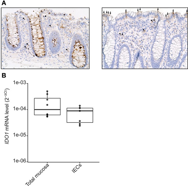
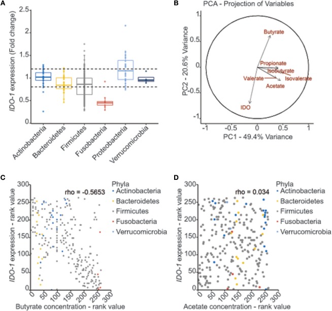
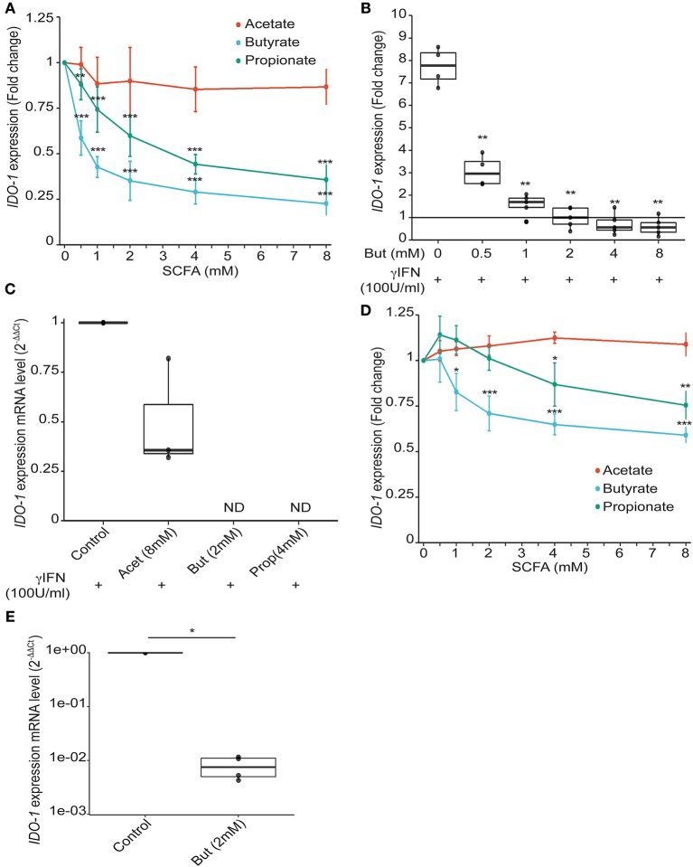
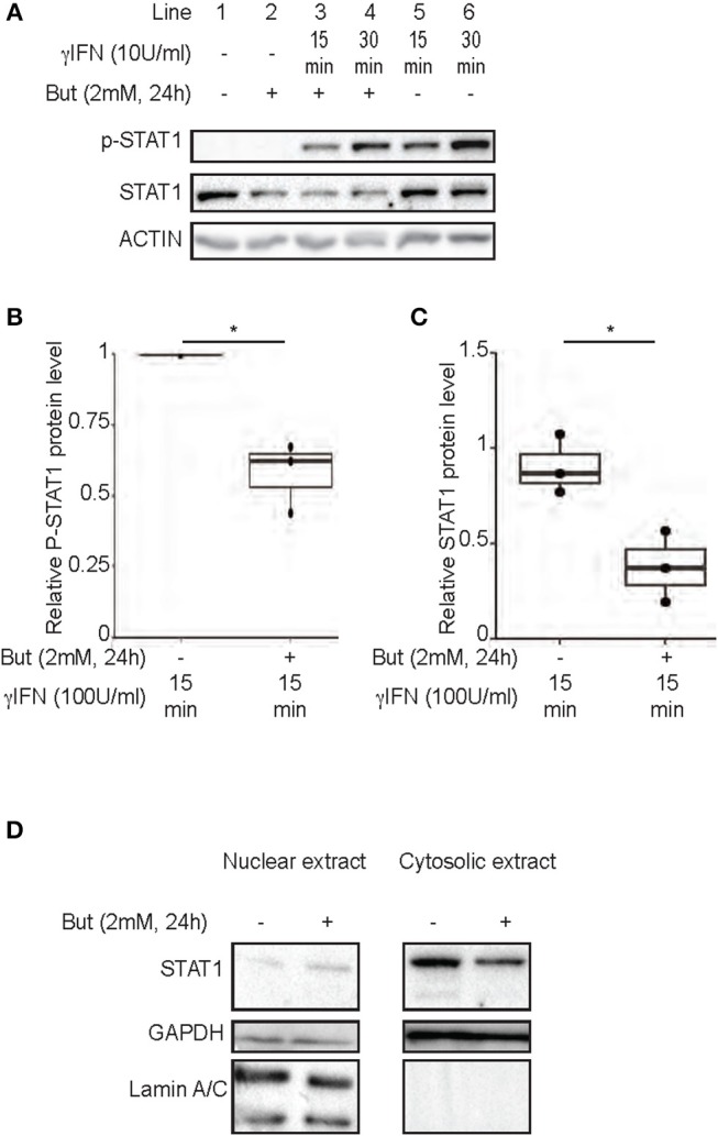
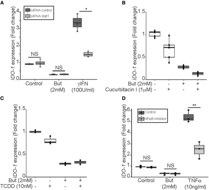
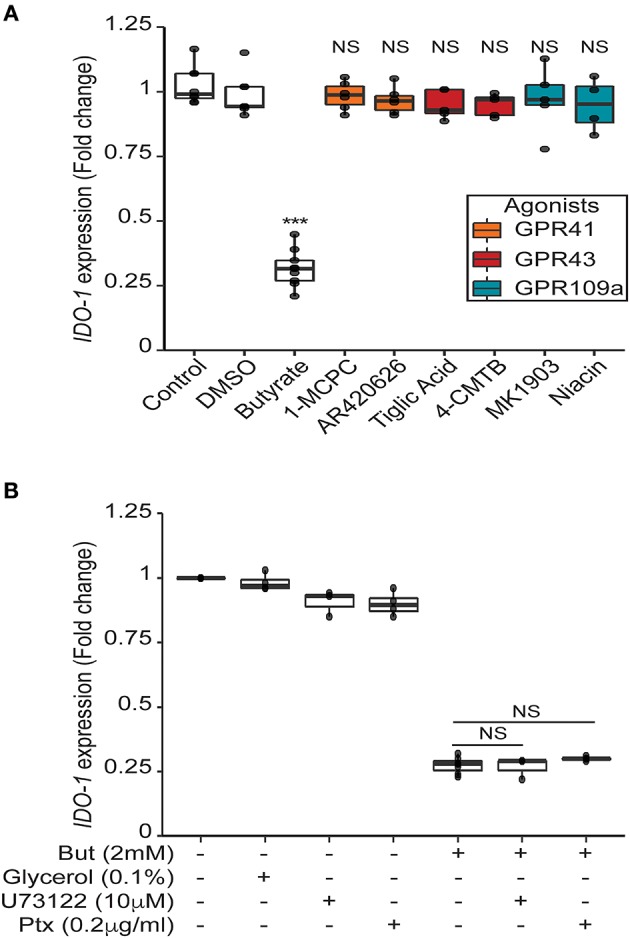
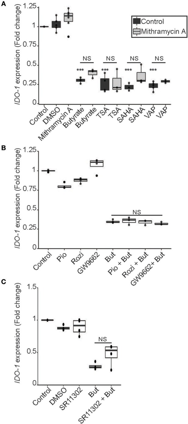
Commensal bacteria are crucial for the development and maintenance of a healthy immune system therefore contributing to the global well-being of their host. A wide variety of metabolites produced by commensal bacteria are influencing host health but the characterization of the multiple molecular mechanisms involved in host-microbiota interactions is still only partially unraveled. The intestinal epithelial cells (IECs) take a central part in the host-microbiota dialogue by inducing the first microbial-derived immune signals. Amongst the numerous effector molecules modulating the immune responses produced by IECs, indoleamine 2,3-dioxygenase-1 (IDO-1) is essential for gut homeostasis. IDO-1 expression is dependent on the microbiota and despites its central role, how the commensal bacteria impacts its expression is still unclear. Therefore, we investigated the impact of individual cultivable commensal bacteria on IDO-1 transcriptional expression and found that the short chain fatty acid (SCFA) butyrate was the main metabolite controlling IDO-1 expression in human primary IECs and IEC cell-lines. This butyrate-driven effect was independent of the G-protein coupled receptors GPR41, GPR43, and GPR109a and of the transcription factors SP1, AP1, and PPARγ for which binding sites were reported in the IDO-1 promoter. We demonstrated for the first time that butyrate represses IDO-1 expression by two distinct mechanisms. Firstly, butyrate decreases STAT1 expression leading to the inhibition of the IFNγ-dependent and phosphoSTAT1-driven transcription of IDO-1. In addition, we described a second mechanism by which butyrate impairs IDO-1 transcription in a STAT1-independent manner that could be attributed to its histone deacetylase (HDAC) inhibitor property. In conclusion, our results showed that IDO-1 expression is down-regulated by butyrate via a dual mechanism: the reduction of STAT1 level and the HDAC inhibitor property of SCFAs.

Keywords: IDO-1; butyrate; gut microbiota; immune gene regulation; intestinal epithelial cells.


Figures

Figure 1
Figure 1
IDO-1 expression in human colonic epithelial cells. (A) Human normal colonic mucosa was stained for IDO-1. Representative immunohistochemical staining of IDO-1 showed that IDO-1 (brown) is expressed in epithelial cells [left: strong perinuclear and/or membrane staining of about 80% of the IECs; right panel: heterogeneous staining of few IECs (arrows)] and in few lamina propria mononuclear cells (arrowheads) and endothelial cells (asterisk) (original magnification × 200). (B). IDO-1 gene expression was determined by RT-PCR on RNA extracted from preparations of isolated human colonic epithelial cells (IECs) and of whole mucosa microdissected from normal colon. Results were normalized to β-2 microglobulin (B2M) and expressed as 2-ΔCt relative value (median ± quartiles) of 4 patients (1–2 samples/patient).
Figure 2
Figure 2
Correlation between bacterial metabolites production and IDO-1 gene expression. (A) Effect of bacterial supernatants on IDO-1 reporter system organized by phylum. Culture supernatants of a wide range of cultivable commensal bacteria were applied on the HT-29-IDO-1 reporter system (10% vol/vol) for 24 h. IDO-1 expression was measured by luciferase activity and expressed as fold increase toward its control: non inoculated growth medium used for each culture. IDO-1 expression profiles upper and lower the dash lines were considered as significantly changed. (B) PCA analysis showing the correlation between the SCFAs concentrations produced by the commensal bacteria and IDO-1 expression. (C,D) Representation of IDO-1 expression correlated to butyrate (C) and acetate (D) concentration in in bacterial cultures classified by rank value. Actinobacteria in blue, Bacteroidetes in yellow, Firmicutes in gray, Fusobacteria in red and Verrucomicrobiea in light blue.
Figure 3
Figure 3
Impact of SCFAs on IDO-1 expression. (A), HT-29-IDO-1 reporter cells were incubated with a range of concentration of acetate, butyrate and propionate (0.5; 1; 2; 4; 8 mM) for 24 h. IDO-1 expression was measured by luciferase activity and expressed as the mean ± SD fold change toward un-stimulated cells (N > 3). (B), HT-29-IDO-1 reporter cells were incubated with IFNγ (100 U/ml) and a range of concentration of butyrate (0.5–8 mM). IDO-1 expression was measured by luciferase activity and expressed as the median ± quartiles of fold change toward un-stimulated cells (N > 3). (C) IDO-1 gene expression on HT-29 exposed for 6 h to IFNγ (100 U/ml) +/– butyrate (2 mM), propionate (4 mM), or acetate (8 mM) was determined by RT-PCR. Results were normalized to GAPDH and expressed as 2-ΔΔ Ct relative to control mean value; ND: not detected (N = 3). (D) Caco2-IDO-1 reporter cells were incubated with a range of concentration of acetate, propionate and butyrate (0.5; 1; 2; 4; 8 mM). IDO-1 expression was measured by luciferase activity and expressed as the mean ± SD fold change toward un-stimulated cells (N > 3). (E) IDO-1 expression level on human colonic epithelial cells treated for 24 h with butyrate compared to non-treated cells from the same patient was determined by RT-PCR. Results are normalized to RPS17 and expressed as 2-ΔΔ Ct relative to control, median ± quartiles (N = 4). P-value: *P < 0.05, **P < 0.005, ***P < 0.001.
Figure 4
Figure 4
Inhibition of IFNγ-induced IDO-1 expression by butyrate is correlated with a decrease of STAT1 protein level. (A–C) HT-29 cells were cultured 24 h with butyrate (But 2 mM) prior IFNγ (100 U/ml) stimulation for 15 (line 3 with butyrate and 5 without butyrate) or 30 min (line 4 with butyrate and 6 without butyrate). The protein level of p-STAT1 Tyr701, STAT1, and Actin were determined by western-blot on total protein extracted. Densitometric quantifications of total P-STAT1 and STAT1 proteins, from 3 independent experiments, were normalized to Actin and expressed as fold change compared to IFN stimulated cells (B) and unstimulated cell (C), respectively, of 3 independent experiments. Data are represented as median ± quartiles. (D) HT-29 cells were incubated 24 h with medium or butyrate (But 2 mM) prior cytoplasmic and nuclear extractions. The protein levels of STAT1, Laminin A/C and GAPDH were assessed in each fraction by western-blot. P-value: *P < 0.05; NS, Non significant.
Figure 5
Figure 5
Butyrate inhibition of IDO-1 promoter activity is STAT1 and STAT3 independent. (A) HT-29-IDO-1 cells were transfected with STAT1 siRNA or control siRNA and incubated with butyrate (But 2 mM) or IFNγ (100 U/ml) for 24 h before measuring IDO-1 level. (B) HT-29-IDO-1 cells were incubated for 2 h with the STAT3 phosphorylation inhibitor (Cucurbitacin I, 1 μM) prior to butyrate (But 2 mM) treatment for total incubation time of 24 h (N = 4). (C) HT-29-IDO-1 cells were incubated with AHR ligand (TCDD 10 nM) +/– butyrate (But 2 mM) for 24 h. Data represented 2 independent experiments (D) HT-29-IDO-1 cells were incubated for 1 h with the NκFB inhibitor, Bay117082 (Bay 40 μM) prior stimulation with butyrate (But 2 mM) or TNFα (10 ng/ml) for 24 h (N = 3). IDO-1 expression was measured by luciferase activity and expressed as median ± quartiles of fold change toward unstimulated cells. Data represented at least three independent experiments. P-value: *P < 0.05, **P < 0.005, NS, Non significant.
Figure 6
Figure 6
Butyrate mediated impact on IDO-1 is independent of its receptors GPR41, GPR43 and GPR109a. (A) HT-29-IDO-1 reporter cells were incubated for 24 h with selective GPR agonists: GPR41: AR420626 (1 μM) and 1-MCPC (1 mM); GPR43: 4-CMTB (1 μM) and Tiglic acid (1 mM); GPR109a: Niacin (1 mM) and MK1903 (1 μM) or with DMSO (vehicle), butyrate (But 2 mM) or Control (RPMI). (B) HT-29-IDO-1 reporter cells were incubated for 24 h with 2 mM butyrate +/– GPRs sub-unit inhibitors: Pertussis toxin (Ptx, 0.2 μg/ml), U73122 (10 μM) or glycerol (vehicle). IDO-1 expression was measured by luciferase activity and expressed as median ± quartiles of fold change toward un-stimulated cells. Data represented at least three independent experiments. P-value: ***P < 0.001, NS, Non significant.
Figure 7
Figure 7
HDAC inhibitor mimicked the butyrate-dependent down-regulation of IDO-1 expression in a SP1, PPARγ and AP-1 independent manner. (A) HT-29-IDO-1 reporter cells were incubated for 24 h with butyrate (But 2 mM), SAHA (5 μM), Trichostatin A (TSA 1 μM) or Valproic acid (VAP 5 mM) ± SP1 inhibitor (Mitramycin A; MitA 0.1 μM). (B), HT-29-IDO-1 reporter cells were stimulated for 24 h with two PPARγ activators: Pioglitazone (Pio 5 μM); Rosiglitazone (Rosi, 10 μM) or the specific PPARγ inhibitor GW9662 (10 μM) ± butyrate (But 2 mM). (C) HT-29-IDO-1 reporter cells were incubated for 24 h with butyrate (But 2mM) and/or the AP1 inhibitor, SR11302 (10 μM). IDO-1 expression was measured by luciferase activity and expressed as median ± quartiles of fold change toward un-stimulated cells. Data represented at least three independent experiments. P-value: ***P < 0.001, NS, Non significant.

References

    1. Sekirov I, Russell SL, Antunes LC, Finlay BB. Gut microbiota in health and disease. Physiol Rev. (2010) 90:859–904. 10.1152/physrev.00045.2009 - DOI - PubMed
    1. Postler TS, Ghosh S. Understanding the holobiont: how microbial metabolites affect human health and shape the immune system. Cell Metab. (2017) 26:110–30. 10.1016/j.cmet.2017.05.008 - DOI - PMC - PubMed
    1. Blacher E, Levy M, Tatirovsky E, Elinav E. Microbiome-modulated metabolites at the interface of host immunity. J Immunol. (2017) 198:572–80. 10.4049/jimmunol.1601247 - DOI - PubMed
    1. Hinnebusch BF, Meng S, Wu JT, Archer SY, Hodin RA. The effects of short-chain fatty acids on human colon cancer cell phenotype are associated with histone hyperacetylation. J Nutr. (2002) 132:1012–7. 10.1093/jn/132.5.1012 - DOI - PubMed
    1. Schilderink R, Verseijden C, de Jonge WJ. Dietary inhibitors of histone deacetylases in intestinal immunity and homeostasis. Front Immunol. (2013) 4:226. 10.3389/fimmu.2013.00226 - DOI - PMC - PubMed

Publication types

MeSH terms