. 2018 Dec 12:9:2940.
doi: 10.3389/fimmu.2018.02940. eCollection 2018.

Overcoming Target Driven Fratricide for T Cell Therapy

Affiliations

Overcoming Target Driven Fratricide for T Cell Therapy

Eytan Breman et al. Front Immunol. .

Abstract

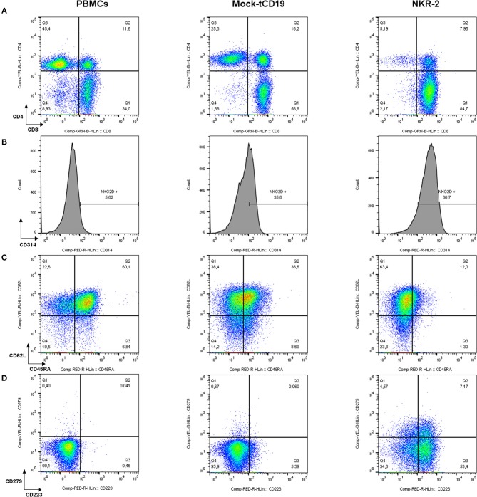
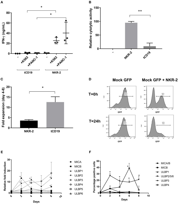
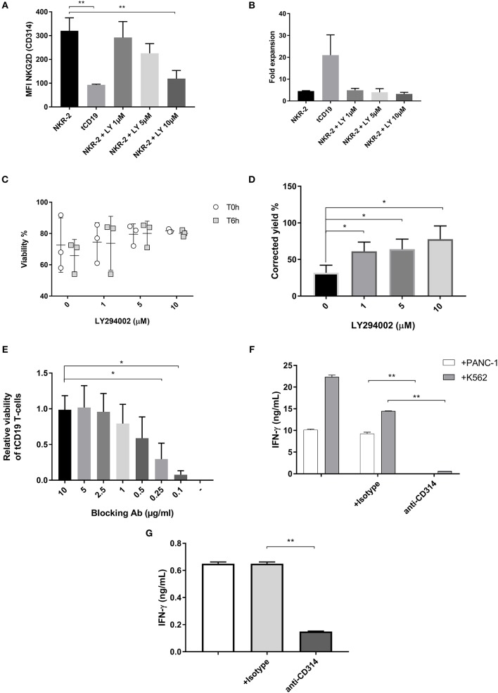
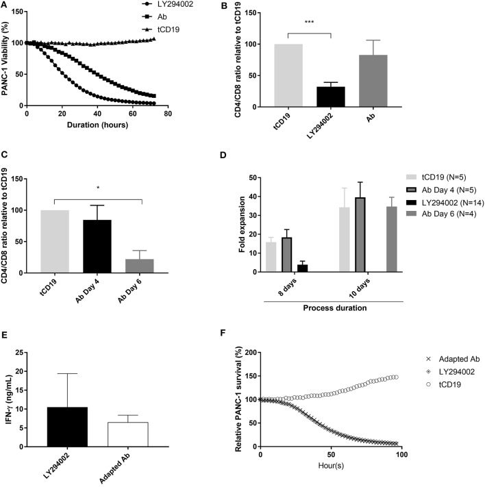
Chimeric Antigen Receptor (CAR) T cells expressing the fusion of the NKG2D protein with CD3ζ (NKG2D-CAR T Cells) acquire a specificity for stress-induced ligands expressed on hematological and solid cancers. However, these stress ligands are also transiently expressed by activated T cells implying that NKG2D-based T cells may undergo self-killing (fratricide) during cell manufacturing or during the freeze thaw cycle prior to infusion in patients. To avoid target-driven fratricide and enable the production of NKG2D-CAR T cells for clinical application, two distinct approaches were investigated. The first focused upon the inclusion of a Phosphoinositol-3-Kinase inhibitor (LY294002) into the production process. A second strategy involved the inclusion of antibody blockade of NKG2D itself. Both processes impacted T cell fratricide, albeit at different levels with the antibody process being the most effective in terms of cell yield. While both approaches generated comparable NKG2D-CAR T cells, there were subtle differences, for example in differentiation status, that were fine-tuned through the phasing of the inhibitor and antibody during culture in order to generate a highly potent NKG2D-CAR T cell product. By means of targeted inhibition of NKG2D expression or generic inhibition of enzyme function, target-driven CAR T fratricide can be overcome. These strategies have been incorporated into on-going clinical trials to enable a highly efficient and reproducible manufacturing process for NKG2D-CAR T cells.

Keywords: CD314; NKG2D; NKG2D blocking antibody; PI3K inhibitor; T cells; chimeric antigen receptor; fratricide.


Figures

Figure 1
Figure 1
Flow cytometric characterization of PBMCs, tCD19, and NKG2D-CAR T cells in a representative NKR-2 T cells process. Mononuclear cells purified from blood using density gradient method were analyzed before (PBMCs) and after (tCD19 and NKR-2 T cells) the NKR-2 T cells process. After acquisition, cells were gated on SSC/FSC for Lymphocytes. Lymphocytes were then gated on CD3. All CD3 positive cells were then displayed as followed: (A) CD4/CD8 distribution. (B) Surface expression of NKG2D (CD314). (C) Memory phenotype. CD62L and CD45RA for the distinction of Naïve (CD62L+ CD45RA+), central memory (CD62L+ CD45RA–), effector memory (CD62L– CD45RA–), and terminal differentiated T cells expressing CD45RA (CD62L– CD45RA+) and (D) Exhaustion phenotype as stained for CD223 (Lag-3) and CD279 (PD-1). One representative donor out of 3 is shown.
Figure 2
Figure 2
NKR-2 T cells recognize Chronic myeloid leukemia (K562) and Pancreatic Carcinoma (PANC-1) but display a fratricide effect due to NKG2DL expression. (A) T cells from healthy donors were transduced with tCD19 vector (tCD19 T cells) or with the NKR-2 T cells vector (NKG2D-CAR T cells) and co-cultured with indicated cell lines or alone (–). IFN-γ secretion (ng/ml) was quantified by ELISA after overnight coculture. Each data point represents the mean value of duplicate wells from independent experiments. Shown is (N = 3). (B) NKR-2 T cells exhibit cytolytic activity. PANC-1 were cocultured with thawed NKG2D-CAR T cells at an E:T ratio of 1:1. Alamar blue signal was determined after 20 h. Percent lysis was determined by absorbance comparison with untreated cancer cells (PANC-1). Data shown are mean ± SD of N = 3 independent T cell donors.; –, PANC-1 cells alone; NKR-2 T cells: Coculture of NKR-2 T cells and PANC-1 cells; tCD19, Coculture of control tCD19 T cells with PANC-1 cells. (C) Transduced T cells were cultured in complete X-vivo (100 IU/mL IL-2) for 4 days. T cells were analyzed 96 h after seeding to analyze fold expansion relative to the initial cell seeding density. Data shown are mean ± SD of N = 3 independent T cell donors. (D) Representative co-culture experiment (out of three). GFP positive T cells were either cultured alone (Mock-GFP) or co-cultured with NKR-2 T cells generated from the same donor (Mock GFP + NKR-2 T cells). Flow cytometry analysis of GFP positivity was acquired at the beginning of the incubation (T = 0 h) or after 24 h of incubation (T = 24 h). (E,F) PBMCs were activated with 40 ng/mL anti-CD3 and 100 IU/mL IL-2 and kept in culture for a total of 8 days in accordance with the normal manufacturing protocol. Every 2 days cell samples were harvested and the expression of NKG2D ligands analyzed as either RNA (E) or proteins on the cell surface (F). A two-tailed unpaired t-test was used to assess statistical significance. A p < 0.05 was considered significant (*) and p < 0.01 (**). For both qPCR and Flow cytometric comparison a paired two-tailed t-test was used.
Figure 3
Figure 3
NKR-2 T cells fratricide is modulated by a PI3K inhibitor or a blocking antibody. (A) MFI of NKG2D expression on NKR-2 T cells treated or not with increasing concentrations of LY294002. Data shown are mean ± SD of N = 3 independent donors. (B) Transduced T cells were cultured in complete X-vivo (100 IU/ml IL-2) supplemented or not with increasing concentrations of LY294002 for expansion. T cells were analyzed 96 h after seeding for fold expansion relative to the initial cell seeding density. Data shown are mean ± SD of N = 3 independent donors. (C) Viability of cells after cryopreservation. NKR-2 T cells were produced in presence of increasing concentration of LY294002. After production, cells were harvested, washed, and formulate for cryopreservation. After cryopreservation, cells were thawed using a water bath and resuspended in Plasmalyte/Human Serum Albumin (HSA) 5%. Cells viability was directly assessed after thawing (T0h) or after 6 h at 4°C in Plasmalyte/HSA5% (T6h) (N = 3) (D) NKR-2 T cells were produced in presence of increasing concentration of LY294002. After production, cells were harvested, washed, transferred in Plasmalyte/HSA1% (50 × 106 NKR-2 T cells/ml) and stored at 4°C for 48 h. After 48 h, cell viability was assessed using trypan blue staining (N = 3) and normalized for the number of cells at cryopreservation (E) tCD19 T cells were co-cultured with NKR-2 T cells generated from the same donor in presence of increasing concentrations of blocking Ab [from 0 (–) to 10 μg/ml]. Flow cytometry analysis of CD19 positivity and viability was acquired after 44 h of incubation. Data are normalized with CD19 positivity of Mock cultured without NKR-2 T cells (N = 3). (F) Thawed NKR-2 T cells were cocultured with either PANC-1 cells or K562 cells at a 1:1 ratio, in the presence of CD314 blocking Ab, an isotype control or no Ab. Following a 24 h incubation, supernatants were harvested and IFN-γ measured (N = 3). (G) Thawed NKR-2 T cells were left in culture for 24h in the presence of an isotype control, the CD314 blocking Ab (or no Ab) and IFN-γ levels measured. Data shown are mean ± SD of N = 3 independent donors. A two-tailed unpaired t-test was used to assess statistical significance. A p < 0.05 was considered significant (*) and **p < 0.01.
Figure 4
Figure 4
NKR-2 T cells Ab blockade process adaptation restores CD4/CD8 ratio. (A) Cytolytic activity kinetics, one representative killing assay out of three. Thawed control tCD19 T cells or NKR-2 T cells treated with PI3K inhibitor LY294002 or with the blocking Ab were cultured in presence of NucLight positive PANC-1. PANC-1 viability was assessed every 2 h by the IncuCyte S3 device. (B) CD4/CD8 distribution at harvest. During expansion phase, NKR-2 T cells were either cultured with 5 μM of LY294002 or with 5 μg/mL of blocking Ab for 96 h, harvested and measured by Flow cytometry for the CD4 and CD8 population. Data shown are mean ± SD of N = 4 independent T cell donors relative to the control tCD19 ratio. (C) CD4/CD8 distribution at harvest with a delayed addition of blocking Ab. During expansion phase (day 4 to day 8), NKR-2 T cells were either directly treated with 5 μM of blocking Ab (day 4) or after 48h (day 6). At harvest (day8), T cells were harvested and analyzed by Flow cytometry for the CD4 and CD8 populations. Data shown are mean ± SD of N = 3 independent T cell donors relative to control tCD19 CD4/CD8 ratio. (D) Comparison of expansion. NKR-2 T cells were either cultured for 8 or 10 days in presence of LY or blocking Ab (added at day 4 or at day 6). T cells were analyzed at harvest for fold expansion relative to the initial cell seeding density. (E) Potency assay by IFN-γ secretion. Thawed NKR-2 T cells processed by the two methods were cocultured in presence of PANC-1 cells. IFN-γ secretion was measured by ELISA after 44 h of co-culture. Data shown are mean ± SD of N = 4 independent T cell donors. (F) Cytolytic activity kinetics, one representative killing assay out of three. Thawed control tCD19 T cells or NKR-2 T cells treated with PI3Ki or with the blocking antibody were cultured in presence of NucLight positive PANC-1 cells. PANC-1 viability was assessed every 2 h by the IncuCyte S3 device. A two-tailed unpaired t-test was used to assess statistical significance. A p < 0.05 was considered significant *p < 0.01 and ***p < 0.001.

References

    1. Fesnak AD, June CH, Levine BL. Engineered T cells: the promise and challenges of cancer immunotherapy. Nat Rev Cancer (2016) 16:566–81. 10.1038/nrc.2016.97 - DOI - PMC - PubMed
    1. Turtle CJ, Hay KA, Hanafi LA, Li D, Cherian S, Chen X, et al. . Durable molecular remissions in chronic lymphocytic leukemia treated with cd19-specific chimeric antigen receptor–modified t cells after failure of ibrutinib. J Clin Oncol. (2017) 35:3010–20. 10.1200/JCO.2017.72.8519 - DOI - PMC - PubMed
    1. Rossig C. CAR T cell immunotherapy in hematology and beyond. Clin Immunol. (2017) 186:54–8. 10.1016/j.clim.2017.09.016 - DOI - PubMed
    1. Park JH, Geyer MB, Brentjens RJ. CD19-targeted CAR T-cell therapeutics for hematologic malignancies: interpreting clinical outcomes to date. Blood (2016) 127:3312–20. 10.1182/blood-2016-02-629063 - DOI - PMC - PubMed
    1. Kochenderfer JN, Somerville RPT, Lu T, Yang JC, Sherry RM, Feldman SA, et al. . Long-Duration complete remissions of diffuse large b cell lymphoma after anti-cd19 chimeric antigen receptor t cell therapy. Mol Ther. (2017) 25:2245–53. 10.1016/j.ymthe.2017.07.004 - DOI - PMC - PubMed

MeSH terms

LinkOut - more resources