. 2018 Dec 20:9:2966.
doi: 10.3389/fimmu.2018.02966. eCollection 2018.

Inhibition of Inflammatory Changes in Human Myometrial Cells by Cell Penetrating Peptide and Small Molecule Inhibitors of NFκB

Affiliations

Inhibition of Inflammatory Changes in Human Myometrial Cells by Cell Penetrating Peptide and Small Molecule Inhibitors of NFκB

Leo R I Gurney et al. Front Immunol. .

Abstract

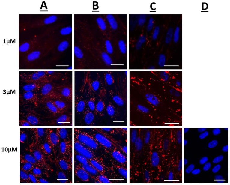
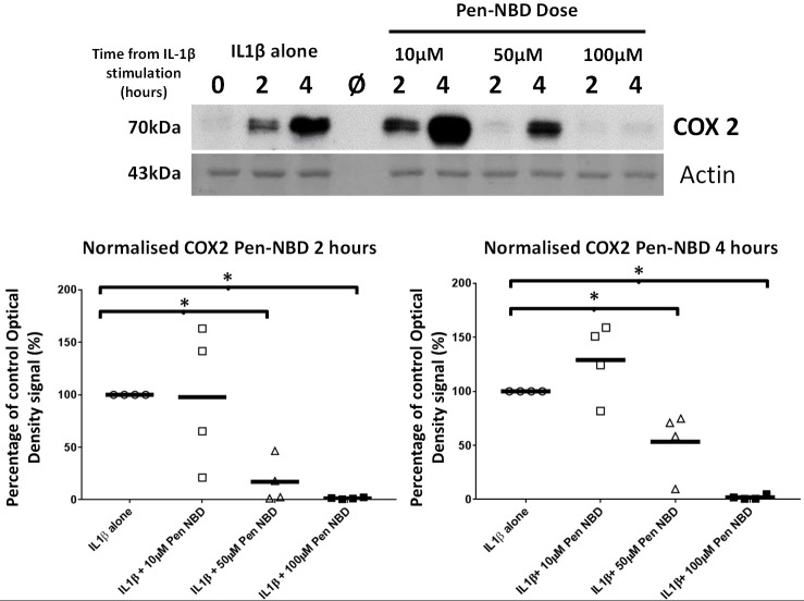
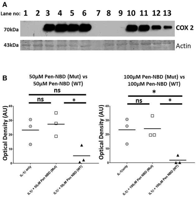
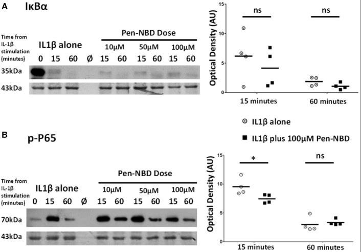
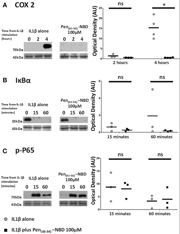
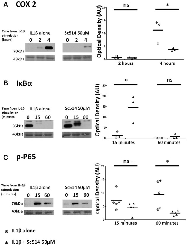
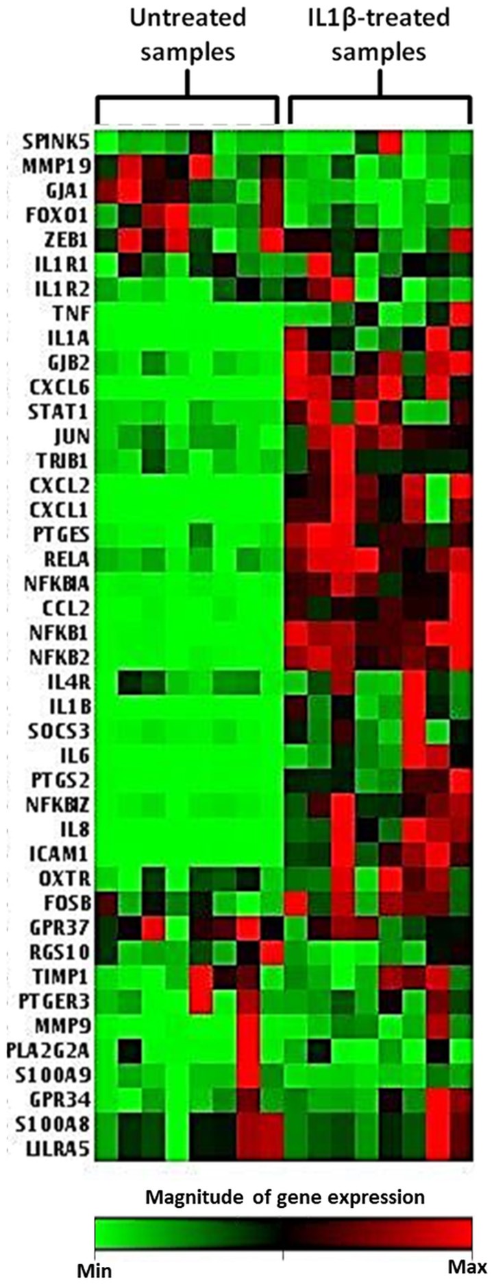
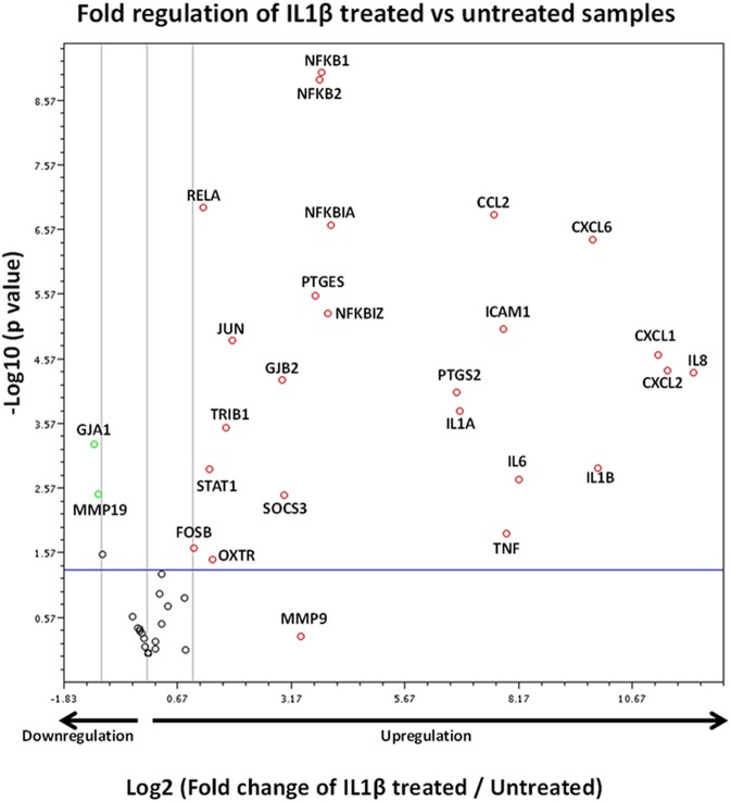
Complications arising from Preterm Birth are the leading causes of neonatal death globally. Current therapeutic strategies to prevent Preterm Birth are yet to demonstrate success in terms of reducing this neonatal disease burden. Upregulation of intracellular inflammatory pathways in uterine cells, including those involving nuclear factor kappa-B (NFκB), have been causally linked to both human term and preterm labor, but the barrier presented by the cell membrane presents an obstacle to interventions aimed at dampening these inflammatory responses. Cell penetrating peptides (CPPs) are novel vectors that can traverse cell membranes without the need for recognition by cell surface receptors and offer the ability to deliver therapeutic cargo internal to cell membranes. Using a human uterine cell culture inflammatory model, this study aimed to test the effectiveness of CPP-cargo delivery to inhibit inflammatory responses, comparing this effect with a small molecule inhibitor (Sc514) that has a similar intracellular target of action within the NFκB pathway (the IKK complex). The CPP Penetratin, conjugated to rhodamine, was able to enter uterine cells within a 60 min timeframe as assessed by live confocal microscopy, this phenomena was not observed with the use of a rhodamine-conjugated inert control peptide (GC(GS)4). Penetratin CPP conjugated to an IKK-inhibitory peptide (Pen-NBD) demonstrated ability to inhibit both the IL1β-induced expression of the inflammatory protein COX2 and dampen the expression of a bespoke array of inflammatory genes. Truncation of the CPP vector rendered the CPP-cargo conjugate much less effective, demonstrating the importance of careful vector selection. The small molecule inhibitor Sc514 also demonstrated ability to inhibit COX2 protein responses and a broad down-regulatory effect on uterine cell inflammatory gene expression. These results support the further exploration of either CPP-based or small molecular treatment strategies to dampen gestational cell inflammatory responses in the context of preterm birth. The work underlines both the importance of careful selection of CPP vector-cargo combinations and basic testing over a broad time and concentration range to ensure effective responses. Further work should demonstrate the effectiveness of CPP-linked cargos to dampen alternative pathways of inflammation linked to Preterm Birth such as MAP Kinase or AP1.

Keywords: cell penetrating peptide; myometrial cell; nuclear factor kappa B; preterm birth; tocolytic.


Figures

Figure 1
Figure 1
Confocal microscopy images of live human uterine smooth muscle cells following application of rhodamine-conjugated CPPs or CPP-cargo combinations. Images captured 60 min following addition of either 1, 3, or 10 μM concentrations of (A) Rhodamine—Penetratin, (B) Rhodamine—Pen-NBD, or (C) Rhodamine—Pen (43-56)-NBD. (D) Displays cells imaged 60 min following addition of 10 μM control peptide Rhodamine - GS4 (GC). Scale bars 20 μm. Cells are maintained in serum deprived media within a temperature-controlled chamber (37°C, 5%CO2). To demonstrate intra-cellularity of uptake, images are taken from the center of a Z-stack of 3–10 slices (0.54 μm apart) intended to capture the full depth of the cell. Images are representative of 3 independent experiments.
Figure 2
Figure 2
Effect of Pen-NBD peptide on IL1β-stimulated COX2 protein increases. (Upper) Representative Western blot demonstrating COX2 protein increase at 2 and 4 h following application of 10 ng/ml IL1β alone or with pre-incubation for 1 h with indicated concentrations of Pen-NBD peptide prior to IL1β addition. Actin expression displayed as loading control. Ø = no protein loaded. (Lower) Scatter plot demonstrating optical density values of COX2 protein signal at 2 and 4 h time points comparing IL1β alone or Pen-NBD plus IL1β experiments. Data presented has been normalized to control signal to demonstrate inhibitory effect. *Significant difference from raw optical density values between IL1β alone and IL1β plus Pen-NBD groups (n = 4, one-way ANOVA with Bonferroni post-hoc correction).
Figure 3
Figure 3
Comparison of effect of Pen-NBD mutant (Mut) peptide with Pen-NBD wild type (WT) on IL1β-stimulated COX2 protein responses. (A) Representative Western blot demonstrating COX2 protein changes at indicated time points. Lanes 1,2,8, and 9 display protein expression with no agonist addition. Lanes 3 and 10 display protein expression with addition of 10 ng/ml IL1β alone. Lanes 4 and 11 display protein expression with DMSO vehicle plus IL1β. Lanes 5 and 6 display protein expression with Pen-NBD (Mut) at indicated concentrations plus IL1β and lanes 12 and 13 display Pen-NBD (WT) at indicated concentrations plus IL1β. Actin expression displayed as loading control. Lane 7—no protein addition. (B) Scatter plots demonstrating comparison of optical density values of COX2 protein signal between IL1β only, IL1β plus Pen-NBD (Mut), or IL1β plus Pen-NBD (WT) at indicated concentrations. *Significant difference from raw optical density values between IL1β only and IL1β plus Pen-NBD groups (n = 3, one-way ANOVA with Bonferroni's post-hoc correction).
Figure 4
Figure 4
Effect of Pen-NBD on the cytokine–induced degradation of IκBα protein and phosphorylation of P65 protein. (Left) Representative Western blots demonstrating responses of (A) IκBα protein and (B) phosphorylated P65 protein at 15 and 60 min following application of 10 ng/ml IL1β either alone or following pre-incubation for 1 h with indicated concentration of Pen-NBD peptide prior to IL1β addition. Actin expression displayed as loading control. Ø = no protein loaded. (Right) Scatter plots demonstrating raw optical density values of (A) IκBα protein and (B) phosphorylated P65 protein signal at 15 and 60 min time points comparing IL1β alone or 100μM Pen-NBD plus IL1β experiments. *Significant difference from raw optical density values between IL1β alone or 100 μM Pen-NBD plus IL1β groups (n = 4, two-way ANOVA with Sidak's post-hoc correction).
Figure 5
Figure 5
Effect of Pen (43-56)-NBD on cytokine-induced alterations of (A) COX2, (B) IκBα, and (C) phosphorylated-P65 proteins. (Left) representative Western blots demonstrating protein responses at indicated time points following application of 10 ng/ml IL1β either alone or following pre-incubation for 1 h with 100 μM of Pen(43-56)-NBD peptide prior to IL1β addition. Actin expression displayed beneath as loading control. (Right) Scatter plots demonstrating protein signal raw optical density values comparing IL1β alone or 100 μM Pen-NBD plus IL1β experiments.*Significant difference from raw optical density values between IL1β alone or 100 μM Pen-NBD plus IL1β groups (n = 3–4, one-way ANOVA with Bonferroni's post-hoc correction).
Figure 6
Figure 6
Effect of Sc514 on cytokine-induced alterations of (A) COX2, (B) IκBα, and (C) phosphorylated-P65 proteins. (Left) representative Western blots demonstrating protein responses at indicated time points following application of 10 ng/ml IL1β either alone or following pre-incubation for 1 h with 50 μM Sc514 prior to IL1β addition. Actin expression displayed beneath as loading control (n = 3 COX2/IκBα, n = 5 p-P65,). (Right) scatter plots demonstrating protein signal raw optical density values comparing IL1β alone or 50 μM Sc514 plus IL1β experiments. *Significant difference from raw optical density values between IL1β alone or 50 μM Sc514 plus IL1β groups (n = 3 COX2/IκBα, n = 5 p-P65 two-way ANOVA with Sidak's post-hoc correction).
Figure 7
Figure 7
Heat map demonstrating myometrial cell gene array expression in response to IL1β cytokine stimulation. The heat map displays gene expression changes from an array of 42 GOI for untreated and IL1β treated samples from eight biological replicates. Highly expressed genes are displayed as shades of red and minimally expressed genes are shown as shades of green (n = 8).
Figure 8
Figure 8
Volcano plot demonstrating gene expression changes in myometrial cells following IL1β exposure. X axis shows fold regulation changes between IL1β treated and untreated samples 4 h following cytokine addition. Y axis is the inverse of the p value generated for each gene of interest (GOI) via students T-test. All genes with >2 fold difference between IL1β treated and untreated samples are labeled on the figure. Upregulated genes are presented as red circles, downregulated genes are green circles and genes demonstrating no change are black circles. Genes above the blue horizontal line represent significant expression changes between IL1β and untreated samples (Student's t-test of the replicate 2ΔΔCt values). Data is summation of experiments from eight biological replicates (n = 8).

References

    1. Blencowe H, Cousens S, Chou D, Oestergaard M, Say LA, Moller B, et al. . Born Too Soon: The global epidemiology of 15 million preterm births. Reprod Health (2013) 10(Suppl. 1): S2. 10.1186/1742-4755-10-S1-S2 - DOI - PMC - PubMed
    1. Costeloe KL, Hennessy EM, Haider S, Stacey F, Marlow N, Draper ES. Short term outcomes after extreme preterm birth in England: comparison of two birth cohorts in 1995 and 2006 (the EPICure studies). BMJ (2012) 345:e7976. 10.1136/bmj.e7976 - DOI - PMC - PubMed
    1. Romero R, Espinoza J, Kusanovic JP, Gotsch F, Hassan S, Erez O, et al. The preterm parturition syndrome. BJOG (2006) 113 (Suppl. 3):17–42. 10.1111/j.1471-0528.2006.01120.x - DOI - PMC - PubMed
    1. Thomson A, Telfer J, Young A, Campbell S, Stewart C, Cameron I, et al. . Leukocytes infiltrate the myometrium during human parturtion: further evidence that labour is an inflammatory process. Hum Reprod. (1999) 14:229–236. 10.1093/humrep/15.1.229 - DOI - PubMed
    1. Osman I, Young A, Ledingham MA, Thomson AJ, Jordan F, Greer IA, Norman JE. Leukocyte density and pro-inflammatory cytokine expression in human fetal membranes, decidua, cervix and myometrium before and during labour at term. Mol. Hum. Reprod. (2003) 9:41–5. 10.1093/molehr/gag001 - DOI - PubMed

Publication types

MeSH terms