. 2018 Dec 19:9:2978.
doi: 10.3389/fimmu.2018.02978. eCollection 2018.

A Mass Spectrometry-Based Profiling of Interactomes of Viral DDB1- and Cullin Ubiquitin Ligase-Binding Proteins Reveals NF-κB Inhibitory Activity of the HIV-2-Encoded Vpx

Affiliations

A Mass Spectrometry-Based Profiling of Interactomes of Viral DDB1- and Cullin Ubiquitin Ligase-Binding Proteins Reveals NF-κB Inhibitory Activity of the HIV-2-Encoded Vpx

Christine D Landsberg et al. Front Immunol. .

Abstract

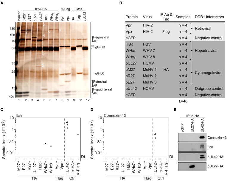
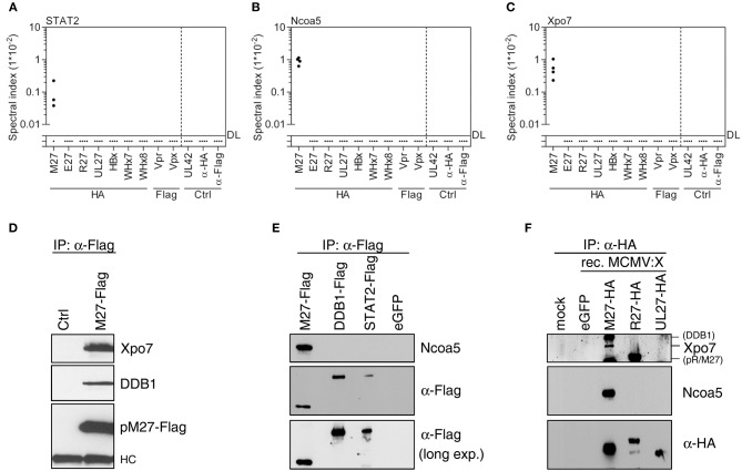
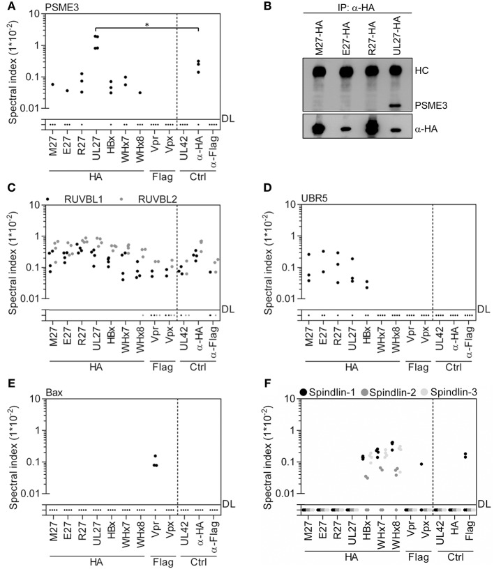
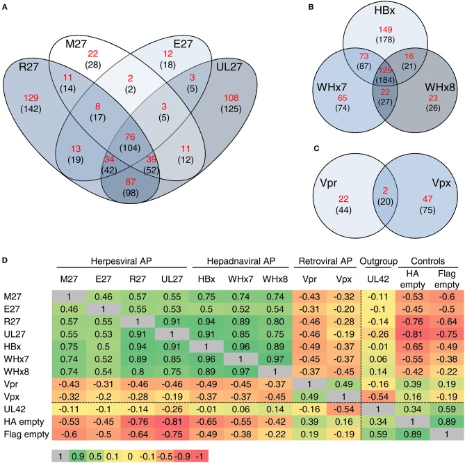
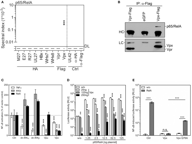
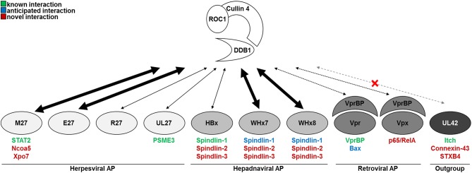
Viruses and hosts are situated in a molecular arms race. To avoid morbidity and mortality, hosts evolved antiviral restriction factors. These restriction factors exert selection pressure on the viruses and drive viral evolution toward increasingly efficient immune antagonists. Numerous viruses exploit cellular DNA damage-binding protein 1 (DDB1)-containing Cullin RocA ubiquitin ligases (CRLs) to induce the ubiquitination and subsequent proteasomal degradation of antiviral factors expressed by their hosts. To establish a comprehensive understanding of the underlying protein interaction networks, we performed immuno-affinity precipitations for a panel of DDB1-interacting proteins derived from viruses such as mouse cytomegalovirus (MCMV, Murid herpesvirus [MuHV] 1), rat cytomegalovirus Maastricht MuHV2, rat cytomegalovirus English MuHV8, human cytomegalovirus (HCMV), hepatitis B virus (HBV), and human immunodeficiency virus (HIV). Cellular interaction partners were identified and quantified by mass spectrometry (MS) and validated by classical biochemistry. The comparative approach enabled us to separate unspecific interactions from specific binding partners and revealed remarkable differences in the strength of interaction with DDB1. Our analysis confirmed several previously described interactions like the interaction of the MCMV-encoded interferon antagonist pM27 with STAT2. We extended known interactions to paralogous proteins like the interaction of the HBV-encoded HBx with different Spindlin proteins and documented interactions for the first time, which explain functional data like the interaction of the HIV-2-encoded Vpr with Bax. Additionally, several novel interactions were identified, such as the association of the HIV-2-encoded Vpx with the transcription factor RelA (also called p65). For the latter interaction, we documented a functional relevance in antagonizing NF-κB-driven gene expression. The mutation of the DDB1 binding interface of Vpx significantly impaired NF-κB inhibition, indicating that Vpx counteracts NF-κB signaling by a DDB1- and CRL-dependent mechanism. In summary, our findings improve the understanding of how viral pathogens hijack cellular DDB1 and CRLs to ensure efficient replication despite the expression of host restriction factors.

Keywords: DNA damage-binding protein (DDB1); NF-κB; cytomegalovirus; hepatitis B virus (HBV); human immunodeficiency virus (HIV); interaction partner; interferon; mass spectrometry (MS).


Figures

Figure 1
Figure 1
Viral DDB1-interacting proteins exhibit distinct co-precipitation profiles. (A) Accessory proteins implicated in the exploitation of DDB1 were expressed in HEK293T cells. Lysates were generated and subjected to IP analysis using the indicated antibodies. Retrieved proteins were visualized by silver staining of SDS gels. Symbols highlight proteins representing partially overlapping and specific interactions. Control IPs were performed using cells transfected with pUL42- and eGFP-expressing plasmids. (B) Overview of the experimental conditions for IP coupled to mass spectrometry (MS). The indicated proteins were analyzed by IP as described in (A) but instead of using silver stained gels, (co-)precipitated proteins were identified and quantified by MS in four biological replicates. Spectral indexes were calculated for Itch (C) and Connexin-43 (D). (E) The indicated proteins were expressed in HEK293T cells. Lysates were subjected to IP using an HA-specific antibody and immunoblot analysis was performed for the indicated proteins.
Figure 2
Figure 2
MCMV-encoded pM27 interacts with STAT2, Xpo7, and Ncoa5. Based on the MS data set (see Figure 1B), spectral indexes were calculated for STAT2 (A), Ncoa5 (B), and Xpo7 (C). (D) NIH3T3 cells were infected (24 h; 5 PFU/cell) with recombinant VACV mutants expressing STAT2-HA (“ctrl”) or pM27-Flag [both described in Zimmermann et al. (14)] to validate the interaction between pM27 and Xpo7. (E) The indicated proteins were expressed in HEK293T cells. Lysates were subjected to IP and subsequent immunoblotting using the indicated antibodies. (F) NIH3T3 cells were infected (24 h; 3 PFU/cell) with recombinant MCMV mutants expressing the indicated cytomegaloviral HA-tagged proteins. The proteins were precipitated using an HA-specific antibody. (Co-) precipitated proteins were analyzed by immunoblotting using the indicated antibodies.
Figure 3
Figure 3
Viral DDB1-interacting accessory proteins co-precipitate DDB1 with distinct efficiencies. Based on the MS data set (see Figure 1B), spectral indexes were calculated. (A) The abundance of DDB1 was calculated as fold over background based on the spectral indexes. (B) The co-precipitation of DDB1 was further analyzed for the herpesviral DDB1-binding proteins by IP and subsequent immunoblotting. The spectral indexes for Cul4B and Cul4A are depicted in (C,D), respectively. (E) The co-precipitation of DDB1 with the hepadnaviral DDB1-binding proteins was analyzed by IP and subsequent immunoblotting. (F) The DDB1 interaction was further analyzed by a reverse approach in which Flag-tagged DDB1 was expressed together with the viral accessory proteins. Lysates were generated, subjected to IP using an α-Flag antibody, and analyzed by immunoblotting.
Figure 4
Figure 4
Exclusive and global interaction partners of viral accessory proteins. Based on the MS data set (see Figure 1B), spectral indexes were calculated. (A) pUL27 precipitated with PSME3. Since PSME3 precipitated to certain extend in the control conditions, the statistical significance was calculated. A level of significance corresponding to p < 0.05 is indicated by “*”. (B) The interaction was confirmed by IP and subsequent immunoblotting. (C) Unspecific precipitation of RUVBL1 and RUVBL2 with viral and control proteins is shown. (D) Herpesviral accessory proteins interact with UBR5. (E) HIV-2 Vpr co-precipitated Bax. (F) Precipitation data for Spindlin-1, -2, and -3 are depicted.
Figure 5
Figure 5
Global comparison of the interactomes of viral DDB1-interacting proteins. Venn diagrams depict the number of interactions shared between the indicated viral proteins. Interaction partners found in at least 2 of 4 replicates were included in the analysis. The calculation was performed for proteins, which were not co-precipitated in any of the control settings (numbers depicted in red) and for all proteins which were at least 2-fold enriched compared to the controls (numbers shown in brackets). The analysis was performed for the herpesviral (A), hepadnaviral (B), and retroviral (C) accessory proteins. (D) For a global comparison, Pearson's correlation coefficients for the entire interactome were calculated and depicted as a heat map including the correlation coefficients. Green color indicates a positive correlation between the interactomes whereas red color depicts an inverse correlation.
Figure 6
Figure 6
HIV-2 Vpx co-precipitates p65/RelA and inhibits NF-κB signaling. (A) Based on the MS data set (see Figure 1B), spectral indexes were calculated for p65/RelA. (B) The interaction was validated by IP and subsequent immunoblotting. Vpr served as control. (C) Cells were co-transfected with an NF-κB-responsive firefly luciferase reporter construct (100 ng), a Gaussia luciferase construct (25 ng) for normalization, and expression vectors for the indicated gene products. NF-κB was activated by TNFα treatment (20 ng/ml) or overexpression of a constitutively active mutant of IKKβ or p65/RelA (both 40 ng). Dominant negative (dn-) IKKα, dn-IKKβ as well as HIV-1 Vpu served as controls (all 100 ng). Luciferase activity was determined 40 h post-transfection. The mean value of 3–9 transfections ± SD is shown. The empty expression plasmid served as control. (D) Increasing concentrations of Vpx and p65/RelA expression plasmids were co-transfected with the NF-κB reporter construct, and luciferase activity was determined at 24 h post-transfection. The empty expression plasmid served as control. The experiment was conducted in n = 3*3 replicates (three independent transfections, each split and measured in triplicates). A level of significance corresponding to p < 0.01 is indicated by “**” whereas p < 0.001 is indicated by “***”. (E) As above, but wt Vpx and a mutant incapable to interact with DDB1 and CRLs (Q76A) were compared. 2.5 ng p65/RelA and 250 ng plasmid encoding Vpx or Vpx-Q76A were transfected. Student's t-Test [two-tailed and heteroscedastic (assuming different variances)] was used to compare the indicated data sets. A level of significance corresponding to p < 0.001 is indicated by “***”.
Figure 7
Figure 7
Model and summary of the findings.

References

    1. Trilling M, Bellora N, Rutkowski AJ, De Graaf M, Dickinson P, Robertson K, et al. . Deciphering the modulation of gene expression by type I and II interferons combining 4sU-tagging, translational arrest and in silico promoter analysis. Nucleic Acids Res. (2013) 41:8107–25. 10.1093/nar/gkt589 - DOI - PMC - PubMed
    1. Megger DA, Philipp J, Le-Trilling VTK, Sitek B, Trilling M. Deciphering of the human interferon-regulated proteome by mass spectrometry-based quantitative analysis reveals extent and dynamics of protein induction and repression. Front Immunol. (2017) 8:1139. 10.3389/fimmu.2017.01139 - DOI - PMC - PubMed
    1. Duggal NK, Emerman M. Evolutionary conflicts between viruses and restriction factors shape immunity. Nat Rev Immunol. (2012) 12:687–95. 10.1038/nri3295 - DOI - PMC - PubMed
    1. Rock KL, Gramm C, Rothstein L, Clark K, Stein R, Dick L, et al. . Inhibitors of the proteasome block the degradation of most cell proteins and the generation of peptides presented on MHC class I molecules. Cell (1994) 78:761–71. - PubMed
    1. Hershko A, Ciechanover A. The ubiquitin system. Ann Rev Biochem. (1998) 67:425–79. - PubMed

Publication types

MeSH terms

Substances