

Ras-Induced miR-146a and 193a Target Jmjd6 to Regulate Melanoma Progression
- PMID: 30619488
- PMCID: PMC6305343
- DOI: 10.3389/fgene.2018.00675
Ras-Induced miR-146a and 193a Target Jmjd6 to Regulate Melanoma Progression
Abstract
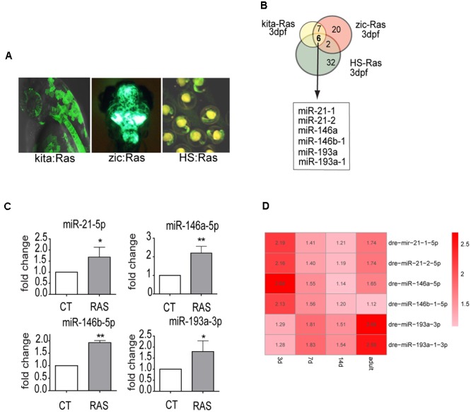
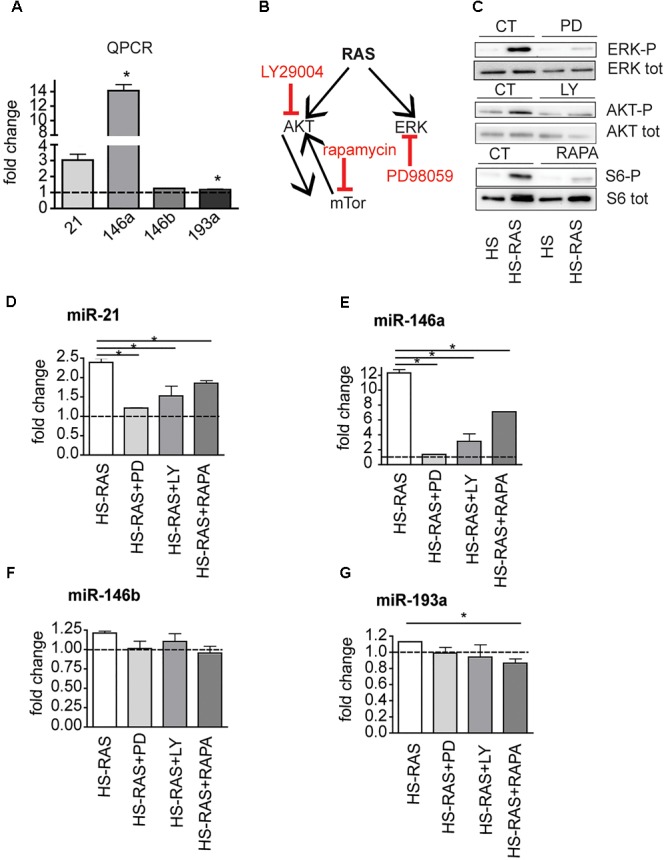
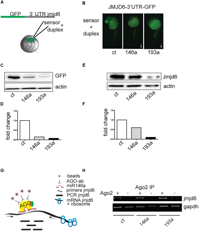
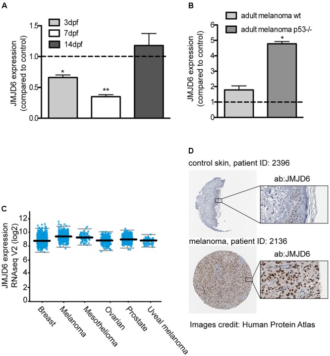
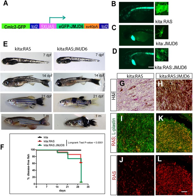
Ras genes are among the most commonly mutated genes in human cancer; yet our understanding of their oncogenic activity at the molecular mechanistic level is incomplete. To identify downstream events that mediate ras-induced cellular transformation in vivo, we analyzed global microRNA expression in three different models of Ras-induction and tumor formation in zebrafish. Six microRNAs were found increased in Ras-induced melanoma, glioma and in an inducible model of ubiquitous Ras expression. The upregulation of the microRNAs depended on the activation of the ERK and AKT pathways and to a lesser extent, on mTOR signaling. Two Ras-induced microRNAs (miR-146a and 193a) target Jmjd6, inducing downregulation of its mRNA and protein levels at the onset of Ras expression during melanoma development. However, at later stages of melanoma progression, jmjd6 levels were found elevated. The dynamic of Jmjd6 levels during progression of melanoma in the zebrafish model suggests that upregulation of the microRNAs targeting Jmjd6 may be part of an anti-cancer response. Indeed, triple transgenic fish engineered to express a microRNA-resistant Jmjd6 from the onset of melanoma have increased tumor burden, higher infiltration of leukocytes and shorter melanoma-free survival. Increased JMJD6 expression is found in several human cancers, including melanoma, suggesting that the up-regulation of Jmjd6 is a critical event in tumor progression. The following link has been created to allow review of record GSE37015: http://www.ncbi.nlm.nih.gov/geo/query/acc.cgi?token=jjcrbiuicyyqgpc&acc=GSE37015.
Keywords: Jmjd6; cancer models; melanoma; miR-146a; miR-193a; microRNA; ras; zebrafish.
Figures
Similar articles
-
MiR-770 inhibits tumorigenesis and EMT by targeting JMJD6 and regulating WNT/β-catenin pathway in non-small cell lung cancer.Life Sci. 2017 Nov 1;188:163-171. doi: 10.1016/j.lfs.2017.09.002. Epub 2017 Sep 4. Life Sci. 2017. PMID: 28882645
-
JMJD6 promotes melanoma carcinogenesis through regulation of the alternative splicing of PAK1, a key MAPK signaling component.Mol Cancer. 2017 Nov 29;16(1):175. doi: 10.1186/s12943-017-0744-2. Mol Cancer. 2017. PMID: 29187213 Free PMC article.
-
Methylation-associated silencing of miR-193a-3p promotes ovarian cancer aggressiveness by targeting GRB7 and MAPK/ERK pathways.Theranostics. 2018 Jan 1;8(2):423-436. doi: 10.7150/thno.22377. eCollection 2018. Theranostics. 2018. PMID: 29290818 Free PMC article.
-
MicroRNA-Based Therapeutic Strategies for Targeting Mutant and Wild Type RAS in Cancer.Drug Dev Res. 2015 Sep;76(6):328-42. doi: 10.1002/ddr.21270. Epub 2015 Aug 18. Drug Dev Res. 2015. PMID: 26284568 Free PMC article. Review.
-
MicroRNA regulation of K-Ras in pancreatic cancer and opportunities for therapeutic intervention.Semin Cancer Biol. 2019 Feb;54:63-71. doi: 10.1016/j.semcancer.2017.11.020. Epub 2017 Dec 2. Semin Cancer Biol. 2019. PMID: 29199014 Free PMC article. Review.
Cited by
-
JMJD family proteins in cancer and inflammation.Signal Transduct Target Ther. 2022 Sep 1;7(1):304. doi: 10.1038/s41392-022-01145-1. Signal Transduct Target Ther. 2022. PMID: 36050314 Free PMC article. Review.
-
Role of the Epigenetic Modifier JMJD6 in Tumor Development and Regulation of Immune Response.Front Immunol. 2022 Mar 14;13:859893. doi: 10.3389/fimmu.2022.859893. eCollection 2022. Front Immunol. 2022. PMID: 35359945 Free PMC article. Review.
-
Zebrafish-based platform for emerging bio-contaminants and virus inactivation research.Sci Total Environ. 2023 May 10;872:162197. doi: 10.1016/j.scitotenv.2023.162197. Epub 2023 Feb 11. Sci Total Environ. 2023. PMID: 36781138 Free PMC article. Review.
-
Craniofacial and Dental Anomalies of a Patient Carrying Two MicroRNA Variants: A Proof-Of-Concept Case Report.Clin Case Rep. 2025 Apr 7;13(4):e70137. doi: 10.1002/ccr3.70137. eCollection 2025 Apr. Clin Case Rep. 2025. PMID: 40201796 Free PMC article.
-
miR-146a-5p impairs melanoma resistance to kinase inhibitors by targeting COX2 and regulating NFkB-mediated inflammatory mediators.Cell Commun Signal. 2020 Sep 23;18(1):156. doi: 10.1186/s12964-020-00601-1. Cell Commun Signal. 2020. PMID: 32967672 Free PMC article.
References
-
- Boeckel J.-N., Guarani V., Koyanagi M., Roexe T., Lengeling A., Schermuly R. T., et al. (2011). Jumonji domain-containing protein 6 (Jmjd6) is required for angiogenic sprouting and regulates splicing of VEGF-receptor 1. Proc. Natl. Acad. Sci. U.S.A. 108 3276–3281. 10.1073/pnas.1008098108 - DOI - PMC - PubMed
-
- Cerami E., Gao J., Dogrusoz U., Gross B. E., Sumer S. O., Aksoy B. A., et al. (2012). The cBio cancer genomics portal: an open platform for exploring multidimensional cancer genomics data. Cancer Discov. 2 401–404. 10.1158/2159-8290.CD-12-0095 - DOI - PMC - PubMed
LinkOut - more resources
Full Text Sources
Molecular Biology Databases
Miscellaneous