Review
. 2018 Dec 13:5:110.
doi: 10.3389/fmolb.2018.00110. eCollection 2018.

D-3-Phosphoglycerate Dehydrogenase

Affiliations
Review

D-3-Phosphoglycerate Dehydrogenase

Gregory A Grant. Front Mol Biosci. .

Abstract

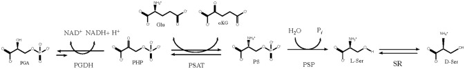
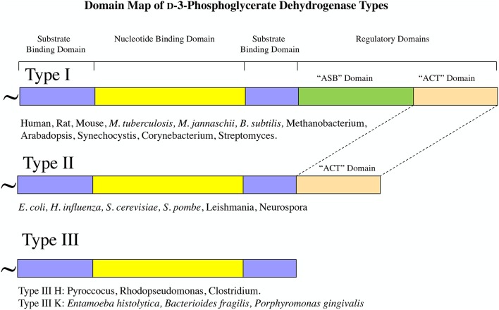
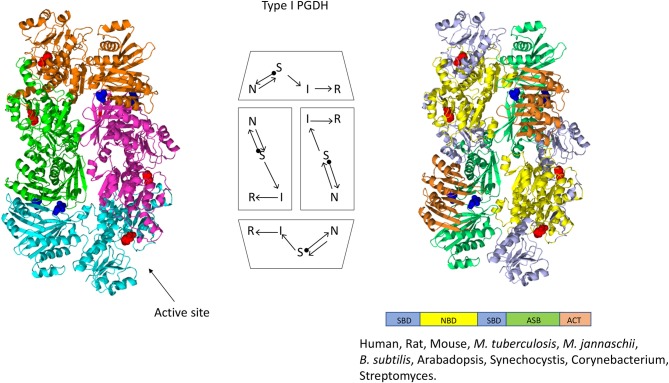
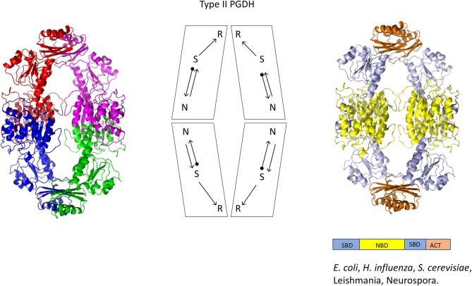
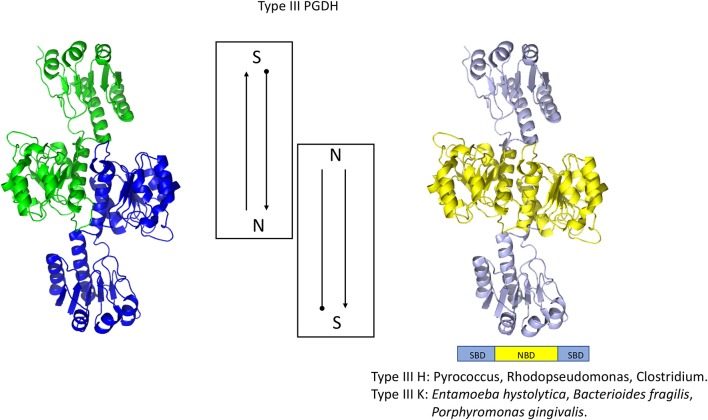
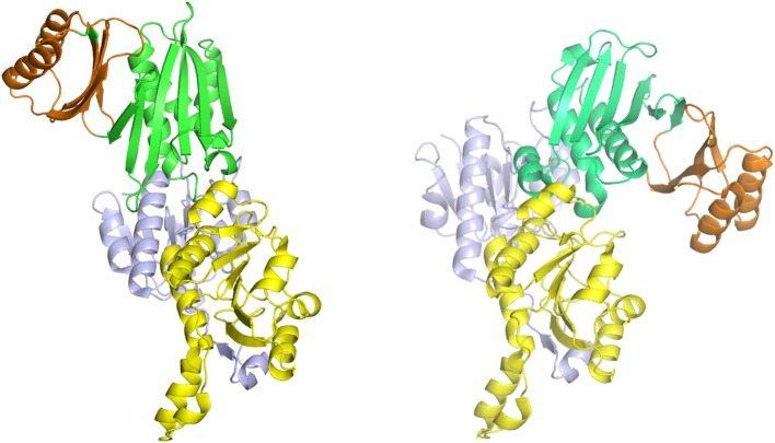
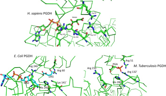
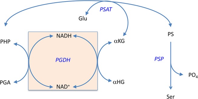
l-Serine is the immediate precursor of d-serine, a major agonist of the N-methyl-d-aspartate (NMDA) receptor. l-Serine is a pivotal amino acid since it serves as a precursor to a large number of essential metabolites besides d-serine. In all non-photosynthetic organisms, including mammals, a major source of l-serine is the phosphorylated pathway of l-serine biosynthesis. The pathway consists of three enzymes, d-3-phosphoglycerate dehydrogenase (PGDH), phosphoserine amino transferase (PSAT), and l-phosphoserine phosphatase (PSP). PGDH catalyzes the first step in the pathway by converting d-3-phosphoglycerate (PGA), an intermediate in glycolysis, to phosphohydroxypyruvate (PHP) concomitant with the reduction of NAD+. In some, but not all organisms, the catalytic activity of PGDH can be regulated by feedback inhibition by l-serine. Three types of PGDH can be distinguished based on their domain structure. Type III PGDHs contain only a nucleotide binding and substrate binding domain. Type II PGDHs contain an additional regulatory domain (ACT domain), and Type I PGDHs contain a fourth domain, termed the ASB domain. There is no consistent pattern of domain content that correlates with organism type, and even when additional domains are present, they are not always functional. PGDH deficiency results in metabolic defects of the nervous system whose systems range from microcephaly at birth, seizures, and psychomotor retardation. Although deficiency of any of the pathway enzymes have similar outcomes, PGDH deficiency is predominant. Dietary or intravenous supplementation with l-serine is effective in controlling seizures but has little effect on psychomotor development. An increase in PGDH levels, due to overexpression, is also associated with a wide array of cancers. In culture, PGDH is required for tumor cell proliferation, but extracellular l-serine is not able to support cell proliferation. This has led to the hypothesis that the pathway is performing some function related to tumor growth other than supplying l-serine. The most well-studied PGDHs are bacterial, primarily from Escherichia coli and Mycobacterium tuberculosis, perhaps because they have been of most interest mechanistically. However, the relatively recent association of PGDH with neuronal defects and human cancers has provoked renewed interest in human PGDH.

Keywords: biosynthesis; d-serine; dehydrogenase; l-serine; phosphoglycerate.


Figures

Figure 1
Figure 1
The l-serine biosynthetic pathway uses d-3 phosphoglycerate from glycolysis in the first step. d-serine is produced from l-serine by serine racemase.
Figure 2
Figure 2
PGDH exists as three different types distinguished by domain structure. The domain structure, going from amino terminus to carboxyl terminus, is illustrated for each type along with representative species containing each type. The “~” symbol signifies that the enzyme from different species can contain different lengths of amino acid sequence at the amino terminus.
Figure 3
Figure 3
Amino acid sequence alignment of PGDH form representative species. Mt, Mycobacterium tuberculosis; Dr, Danio rerio (zebrafish); Rn, Rattus norvegicus; Hs, Homo sapiens; Sc1 and Sc2, two variants from Saccharomyces cervisiae; Ec, Escherichia coli; Eh, Entamoeba hystolytica; Dm, Drosophila melanogaster. The numbering designates residue position in the figure rather than the sequence of a particular PGDH. The PGDH type is shown as a numeral in front of the species abbreviation. The domains are highlighted with colored lines. Substrate binding domain, blue; nucleotide binding domain, yellow; ASB domain, green; and ACT domain, orange. Conserved residues are highlighted in yellow. Residues involved in l-serine binding at the ACT site are highlighted in green. The active site lysine in some type 3 enzymes is highlighted in magenta. Asterisks designate residues involved in substrate and effector binding and the plus sign identifies the active site histidine or lysine. The carboxyl termini are designated “Ct”.
Figure 4
Figure 4
Crystal structure of a Type I PGDH from M. tuberculosis. On the left, each subunit is colored differently. In the center is a diagram of the subunits showing the course of the polypeptide chain from amino terminus, designated by a black dot, to the carboxyl terminus. On the right, the individual domains are colored according to the scheme shown below the structure. Representative species with Type 1 enzymes are listed.
Figure 5
Figure 5
Crystal structure of a Type II PGDH from E. coli. On the left, each subunit is colored differently. In the center is a diagram of the subunits showing the course of the polypeptide chain from amino terminus, designated by a black dot, to the carboxyl terminus. On the right, the individual domains are colored according to the scheme shown below the structure. Representative species with Type II enzymes are listed.
Figure 6
Figure 6
Crystal structure of a Type III PGDH from E. hystolytica. On the left, each subunit is colored differently. In the center is a diagram of the subunits showing the course of the polypeptide chain from amino terminus, designated by a black dot, to the carboxyl terminus. On the right, the individual domains are colored according to the scheme shown below the structure. Representative species with Type III enzymes are listed.
Figure 7
Figure 7
The syn- (right) and anti- (left) configurations of the subunits of M. tuberculosis PGDH. The subunits are pictured with their substrate (blue) and nucleotide (yellow) binding domains in essentially the same orientation. The ASB (green) and ACT (orange) domains are rotated to the left in the anti-configuration and to the right in the syn-configuration.
Figure 8
Figure 8
(Top) Depiction of the active site of human PGDH from pdb 2g76 with malate and NAD bound. Some of the alpha chain in the background has been cut away for clarity. (Bottom) Depiction of the active site of E. coli PGDH from pdb 1yba (left) and of M. tuberculosis PGDH from pdb 2ddn (right).
Figure 9
Figure 9
(Left) The ACT domain interface in E. coli PGDH showing the interaction of l-serine with specific residues. Reprinted from Grant (2012). (Top Right) The serine binding site in E. coli PGDH. The serine atoms are shown as spheres in the right-hand structure. Reprinted from Grant (2012), with permission from Elsevier © 2011. (Bottom Left) The serine binding site in M. tuberculosis PGDH. The serine atoms are shown as spheres in the left-hand structure. Reprinted from Grant (2012), with permission from Elsevier © 2011.
Figure 10
Figure 10
The relationship of the ACT (orange) and ASB (green) domains in M. tuberculosis PGDH. The depiction on the left shows the domains in the absence of bound ligands and the depiction on the right shows L-serine and tartrate bound at their respective sites. The stick model at the bottom shows the specific residue interactions seen in the crystal structure.
Figure 11
Figure 11
The self-sustaining cycle that regenerates coenzyme bound to PGDH during biosynthesis of l-serine in E. coli. The NADH that is produced during the conversion of PGA to PHP is converted back to NAD+ in situ by conversion of αKG to αHG. The αKG is formed from glutamate by the second enzyme in the pathway, PSAT. Reprinted from Grant (2018) with permission from the American Chemical Society.

References

    1. Abe T., Suzuki M., Sasabe J., Takahashi S., Unekawa M., Mashima K., et al. . (2014). Cellular origin and regulation of D- and L-serine in in vitro and in vivo models of cerebral ischemia. J. Cell. Blood Flow Metab. 34, 1928–1935. 10.1038/jcbfm.2014.164 - DOI - PMC - PubMed
    1. Achouri Y., Rider M. H., Van Schaftingen E., Robbi M. (1997). Cloning, sequencing and expression of rat liver 3-phosphoglycerate dehydrogenase. Biochem. J. 323, 365–370. 10.1042/bj3230365 - DOI - PMC - PubMed
    1. Ali V., Hashimoto T., Shigeta Y., Nozaki T. (2004). Molecular and biochemical characterization of d-phosphoglycerate dehydrogenase from Entamoeba histolytica: a unique enteric protozoan parasite that possesses both phosphorylated and nonphosphorylated serine metabolic pathways. Eur. J. Biochem. 271, 2670–2681. 10.1111/j.1432-1033.2004.04195.x - DOI - PubMed
    1. Al-Rabiee R., Lee E. J., Grant G. A. (1996a). The mechanism of velocity modulated allosteric regulation in d-3-Phosphoglycerate dehydrogenase: cross-linking adjacent regulatory domains with engineered disulfides mimics effector binding. J. Biol. Chem. 271, 13013–13017. 10.1074/jbc.271.22.13013 - DOI - PubMed
    1. Al-Rabiee R., Zhang Y., Grant G. A. (1996b). The mechanism of velocity modulated allosteric regulation in d-3-phosphoglycerate dehydrogenase. Site directed mutagenesis of effector binding sites. J. Biol. Chem. 271, 23235–23238. 10.1074/jbc.271.38.23235 - DOI - PubMed

LinkOut - more resources