

Evaluation of cerebral blood flow in older patients with status epilepticus using arterial spin labeling
- PMID: 30619954
- PMCID: PMC6313842
- DOI: 10.1016/j.ensci.2018.12.005
Evaluation of cerebral blood flow in older patients with status epilepticus using arterial spin labeling
Abstract
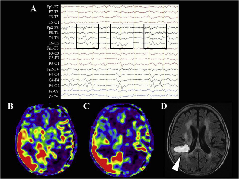
Introduction: Although older patients with status epilepticus (SE) have a high mortality rate and poor outcome, it is difficult to perform emergent electroencephalography (EEG) to diagnose SE in community hospitals. Arterial spin labeling (ASL) is a non-invasive magnetic resonance imaging (MRI) technique that can rapidly assess cerebral blood flow (CBF). Further, ASL can detect increased CBF in the ictal period. Therefore, ASL may be a useful tool for diagnosing SE in older patients. However, its effectiveness in this population is unknown.
Methods: We retrospectively investigated differences in CBF abnormalities between older patients (≥70 years) and non-older patients (<70 years) with SE using ASL. Participants were diagnosed with convulsive status epilepticus (CSE) or non-convulsive status epilepticus (NCSE) based on symptoms, brain MRI, and EEG.
Results: ASL detected CBF abnormalities in 40% of older patients with CSE or NCSE. Rates of CBF abnormalities in older patients were not significantly different compared with that in non-older patients.
Conclusions: ASL did not detect a higher rate of CBF abnormalities in older patients, but may help physicians diagnose SE in older patients in a community hospital setting if emergent EEG cannot be immediately performed.
Keywords: Arterial spin labeling; Magnetic resonance imaging; Non-convulsive status epilepticus; Older patient; Status epilepticus.
Figures
References
-
- Trinka E., Cock H., Hesdorffer D. A definition and classification of status epilepticus—report of the ILAE task force on classification of status epilepticus. Epilepsia. 2015;56(10):1515–1523. - PubMed
-
- Leitinger M., Beniczky S., Rohracher A. Salzburg consensus criteria for non-convulsive status epilepticus—approach to clinical application. Epilepsy Behav. 2015;49:158–163. - PubMed
-
- Varelas P.N., Spanaki M.V., Hacein-Bey L. Emergent EEG: indications and diagnostic yield. Neurology. 2003;61(5):702–704. - PubMed
LinkOut - more resources
Full Text Sources
