

Case Reports
doi: 10.1016/j.jvscit.2018.09.006.
eCollection 2019 Mar.
Rotational vertebral artery occlusion secondary to completely extraosseous vertebral artery
Affiliations
- PMID: 30619984
- PMCID: PMC6313838
- DOI: 10.1016/j.jvscit.2018.09.006
Item in Clipboard
Case Reports
Rotational vertebral artery occlusion secondary to completely extraosseous vertebral artery
J Vasc Surg Cases Innov Tech.
.
Abstract
Rotational vertebral artery (VA) occlusion is a possible cause of reduced blood flow through the posterior circulation of the brain due to compression of the VA on head turning when blood flow from the contralateral VA is compromised. When compression occurs in the V2 segment of the VA, it is usually due to compression from the longus colli muscle or cervical osteophytes. We present a unique case of a patient with a completely extraosseous course of the V2 segment of her dominant right VA that resulted in symptomatic rotational VA occlusion.
Keywords: Compression; Syncope; Vertebral artery; Vertebrobasilar insufficiency.
Figures
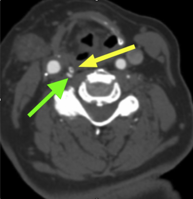
Preoperative computed tomography angiography (CTA) demonstrates extrinsic compression of the extraosseous V2 segment of the right vertebral artery (VA). The yellow arrow points to the posterior superior projection of the thyroid cartilage. The green arrow demonstrates extrinsic compression of the right VA by the posterior superior projection of the thyroid cartilage.
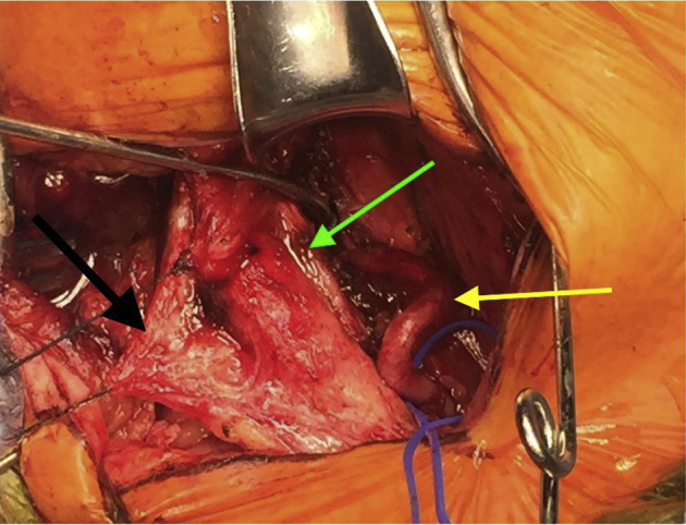
The yellow arrow points to the extraosseous right vertebral artery (VA) and the green arrow shows the levator scapulae muscle coursing anterior to and compressing the VA. The black arrow points to the posterior carotid sheath being retracted with stay sutures. The patient's head is in a neutral position.
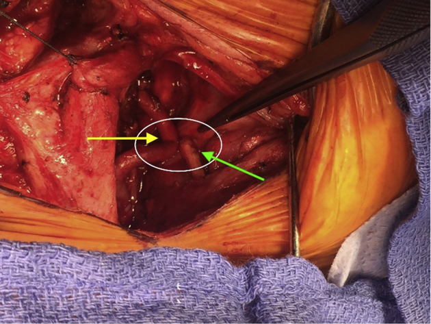
The extraosseous V2 segment of the right vertebral artery (VA) is demonstrated by the yellow arrow. The green arrow points to the posterior superior projection of the right thyroid cartilage. The area of extrinsic compression of the right VA is circled in white.
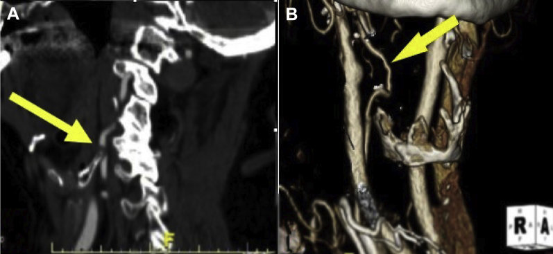
Comparison of the preoperative (A) and postoperative (B) volume rendered computed tomography angiograms of the vertebral artery (VA; arrows) demonstrates that segmental removal of the thyroid cartilage has resolved the rotationally induced compression.
Similar articles
-
Asymptomatic rotational vertebral artery compression in a child due to head positioning for cranial surgery: illustrative case.J Neurosurg Case Lessons. 2021 Jan 18;1(3):CASE2085. doi: 10.3171/CASE2085. eCollection 2021 Jan 18. J Neurosurg Case Lessons. 2021. PMID: 36034509 Free PMC article.
-
Rotational vertebrobasilar insufficiency due to compression of a persistent first intersegmental vertebral artery variant: case report.J Neurosurg Spine. 2017 Feb;26(2):199-202. doi: 10.3171/2016.7.SPINE163. Epub 2016 Oct 7. J Neurosurg Spine. 2017. PMID: 27716015
-
Rotational vertebral artery occlusion secondary to adjacent-level degeneration following anterior cervical discectomy and fusion.J Neurosurg Spine. 2014 Jun;20(6):714-21. doi: 10.3171/2014.3.SPINE13452. Epub 2014 Apr 18. J Neurosurg Spine. 2014. PMID: 24745352
-
Rotational vertebral artery occlusion: a mechanism of vertebrobasilar insufficiency.Neurosurgery. 1997 Aug;41(2):427-32; discussion 432-3. doi: 10.1097/00006123-199708000-00019. Neurosurgery. 1997. PMID: 9257311 Review.
-
Bow Hunter's Syndrome by Nondominant Vertebral Artery Compression: A Case Report, Literature Review, and Significance of Downbeat Nystagmus as the Diagnostic Clue.World Neurosurg. 2018 Mar;111:367-372. doi: 10.1016/j.wneu.2017.12.167. Epub 2018 Jan 5. World Neurosurg. 2018. PMID: 29309982 Review.
Cited by
-
Subaxial Vertebral Artery Rotational Occlusion Syndrome: An Overview of Clinical Aspects, Diagnostic Work-Up, and Surgical Management.Asian Spine J. 2021 Jun;15(3):392-407. doi: 10.31616/asj.2020.0275. Epub 2020 Sep 10. Asian Spine J. 2021. PMID: 32898967 Free PMC article.
-
Vertigo in Patients with Degenerative Cervical Myelopathy.J Clin Med. 2021 Jun 4;10(11):2496. doi: 10.3390/jcm10112496. J Clin Med. 2021. PMID: 34200086 Free PMC article.
-
Atypical presentation of rotational vertebral artery insufficiency: illustrative case.J Neurosurg Case Lessons. 2021 Mar 1;1(9):CASE20169. doi: 10.3171/CASE20169. eCollection 2021 Mar 1. J Neurosurg Case Lessons. 2021. PMID: 35854706 Free PMC article.
-
Bow hunter's syndrome due to an anomalous right vertebral artery origin and contralateral absence: a case report and literature review.BMC Neurol. 2024 Jul 12;24(1):242. doi: 10.1186/s12883-024-03754-5. BMC Neurol. 2024. PMID: 38997640 Free PMC article. Review.
-
Recurrent infarcts from thyroid cartilage compression of an aberrant vertebral artery: rare, easily overlooked, but treatable.J Neurol. 2023 Dec;270(12):6146-6150. doi: 10.1007/s00415-023-11896-8. Epub 2023 Aug 7. J Neurol. 2023. PMID: 37548680 Free PMC article. No abstract available.
References
-
- Pearl G.J., Shutze W.P. Vertebral artery disease. In: Valentine R.J., Eidt J.F., editors. Scientific American vascular and endovascular surgery [online] Decker Intellectual Properties; Hamilton, Ontario: 2015.
-
- Lee V., Riles T.S., Stableford J., Berguer R. Two case presentations and surgical management of Bow Hunter's syndrome associated with bony abnormalities of the C7 vertebra. J Vasc Surg. 2011;53:1381–1385. - PubMed
-
- Kuether T.A., Nesbit G.M., Clark W.M., Barnwell S.L. Rotational vertebral artery occlusion: a mechanism of vertebrobasilar insufficiency. Neurosurgery. 1997;41:427–432. discussion: 432-3. - PubMed
-
- Lee C.S., Lee H.Y., Yang T.K. Rotational vertebral artery occlusion syndrome: misnomers and classification. Clin Neurol Neurosurg. 2015;131:18–20. - PubMed
-
- Shutze W., Gierman J., McQuade K., Pearl G., Smith B. Treatment of proximal vertebral artery disease. Vascular. 2014;22:85–92. - PubMed
Publication types
LinkOut - more resources
Full Text Sources
