. 2018 Oct 16;3(1):181-200.
doi: 10.1210/js.2018-00226. eCollection 2019 Jan 1.

Pituitary Pathology and Gene Expression in Acromegalic Cats

Affiliations

Pituitary Pathology and Gene Expression in Acromegalic Cats

Christopher J Scudder et al. J Endocr Soc. .

Abstract

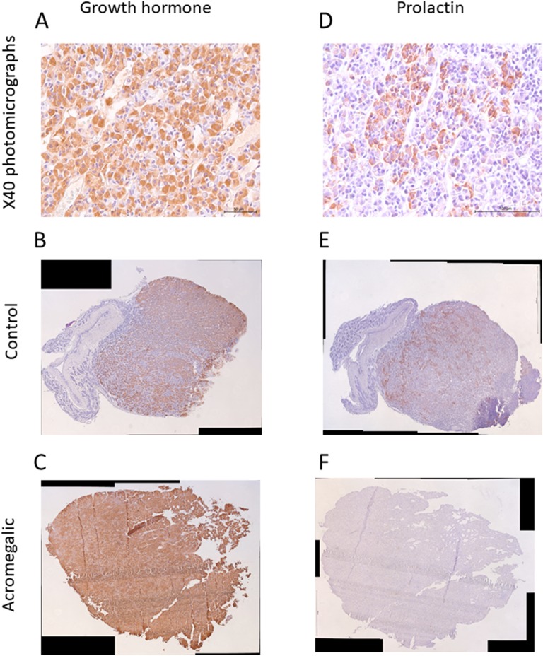
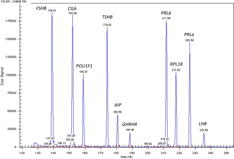
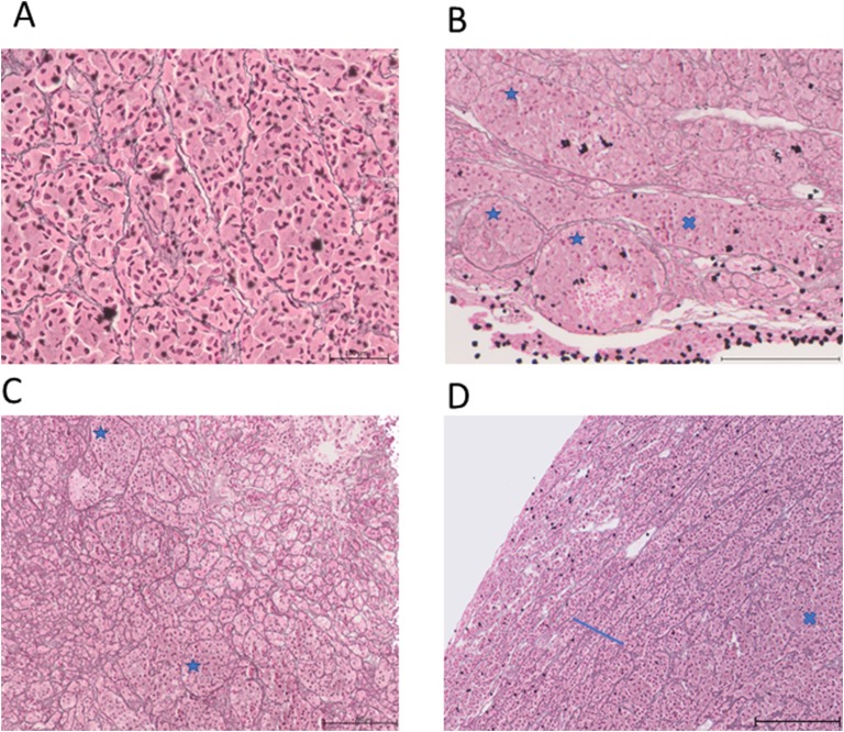
The prevalence of GH-secreting pituitary tumors in domestic cats (Felis catus) is 10-fold greater than in humans. The predominant inhibitory receptors of GH-secreting pituitary tumors are somatostatin receptors (SSTRs) and D2 dopamine receptor (DRD2). The expression of these receptors is associated with the response to somatostatin analog and dopamine agonist treatment in human patients with acromegaly. The aim of this study was to describe pathological features of pituitaries from domestic cats with acromegaly, pituitary receptor expression, and investigate correlates with clinical data, including pituitary volume, time since diagnosis of diabetes, insulin requirement, and serum IGF1 concentration. Loss of reticulin structure was identified in 15 of 21 pituitaries, of which 10 of 15 exhibited acinar hyperplasia. SSTR1, SSTR2, SSTR5, and DRD2 mRNA were identified in the feline pituitary whereas SSTR3 and SSTR4 were not. Expression of SSTR1, SSTR2, and SSTR5 was greater in acromegalic cats compared with controls. A negative correlation was identified between DRD2 mRNA expression and pituitary volume. The loss of DRD2 expression should be investigated as a mechanism allowing the development of larger pituitary tumors.

Keywords: acromegaly; cat; dopamine receptor; hypersomatotropism; somatostatin.


Figures

Figure 1.
Figure 1.
Representative photomicrographs of GH (A–C) and PRL (D–F) immunostaining. (A and D) Immunoreactivity is identified by diaminobenzidine (DAB) chromogen and tissue counterstaining performed using Gill hematoxylin. Photomicrographs demonstrating specific immunostaining for somatrophs and lactotrophs, respectively. Original magnification, ×40 (B and E). Photomicrographs of B–C and E–F were created by obtaining high-resolution photomicrographs at magnification ×100 and stitching images from each tissue together using image editing software. (B and E) Photomicrographs from a control cat. (C and F) Photomicrographs from an acromegalic cat.
Figure 2.
Figure 2.
Representative images of SSTR2 immunoreactivity using feline pituitary tissue. (A–D) Immunoreactivity is identified by diaminobenzidine (DAB) chromogen and tissue counterstaining performed using Gill hematoxylin. Pituitary tissue exhibiting SSTR2 immunohistochemistry scores 0, 1, 2 and 3, respectively, using the following criteria: 0, absent; 1, cytoplasmic staining; 2, membranous staining in <50% cells or incomplete membranous staining; and 3, circumferential membranous staining in >50% cells. All presented photomicrographs were collected at an original magnification of ×100.
Figure 3.
Figure 3.
Electropherogram results from PCR products using multiplex 1 primer sets. The blue peaks represent PCR products from gene-specific primers and the red peaks represent product size standards.
Figure 4.
Figure 4.
Images stained using silver stain for reticulin fibers and counterstained using nuclear fast red solution. (A and C) Reconstructed stitched pituitary photomicrographs from two control pituitaries. Original magnification, ×100. (B and D) Enlarged photomicrographs from sections of (A) and (C), respectively. Original magnification, ×400. The acinar pattern of reticulin staining is identified in (B) and (D). This pattern of reticulin staining was demonstrated in all reticulin staining control pituitaries.
Figure 5.
Figure 5.
All images stained using silver stain for reticulin fibers and counterstained using nuclear fast red solution. (A–D) Selected images taken from reconstructed stitched pituitary photomicrographs from four HST pituitaries. Original magnification, ×100. (A) Disrupted reticulin staining and loss of acinar structure. (B) Areas of enlarged acini (blue stars) and areas of loss of acinar structure (blue cross). (C) Enlarged acini (blue stars) adjacent to normal size and small acini. (D) Loss of acinar structure in the bottom right of the image (blue star); adenomatous tissue has compressed the normal pituitary tissue, resulting in compression of the acini and a ring of cords of acini giving the impression of a pseudocapsule.
Figure 6.
Figure 6.
(A) Bar charts comparing the relative gene expression of SSTR1, SSTR2, and SSTR5 in pituitary tissue from control (CTRL) and acromegalic (Acro) cats determined using the GeXP multiplex technique. RPL18 is the reference gene. Bar height represents mean, and error bars are 95% CIs. **P < 0.01, ***P < 0.001. (B) Dot plot of the individual somatostatin profiles from each of the 19 acromegalic cats.

References

    1. Scacchi M, Cavagnini F. Acromegaly. Pituitary. 2006;9(4):297–303. - PubMed
    1. Giustina A, Chanson P, Kleinberg D, Bronstein MD, Clemmons DR, Klibanski A, van der Lely AJ, Strasburger CJ, Lamberts SW, Ho KK, Casanueva FF, Melmed S; Acromegaly Consensus Group . Expert consensus document: a consensus on the medical treatment of acromegaly. Nat Rev Endocrinol. 2014;10(4):243–248. - PubMed
    1. Katznelson L, Laws ER Jr, Melmed S, Molitch ME, Murad MH, Utz A, Wass JA; Endocrine Society . Acromegaly: an endocrine society clinical practice guideline. J Clin Endocrinol Metab. 2014;99(11):3933–3951. - PubMed
    1. Colao A, Bronstein MD, Freda P, Gu F, Shen CC, Gadelha M, Fleseriu M, van der Lely AJ, Farrall AJ, Hermosillo Reséndiz K, Ruffin M, Chen Y, Sheppard M; Pasireotide C2305 Study Group . Pasireotide versus octreotide in acromegaly: a head-to-head superiority study. J Clin Endocrinol Metab. 2014;99(3):791–799. - PMC - PubMed
    1. Colao A, Auriemma RS, Lombardi G, Pivonello R. Resistance to somatostatin analogs in acromegaly. Endocr Rev. 2011;32(2):247–271. - PubMed

LinkOut - more resources