. 2018 Oct 30;3(1):201-221.
doi: 10.1210/js.2018-00130. eCollection 2019 Jan 1.

Predicted Benign and Synonymous Variants in CYP11A1 Cause Primary Adrenal Insufficiency Through Missplicing

Affiliations

Predicted Benign and Synonymous Variants in CYP11A1 Cause Primary Adrenal Insufficiency Through Missplicing

Avinaash Maharaj et al. J Endocr Soc. .

Abstract

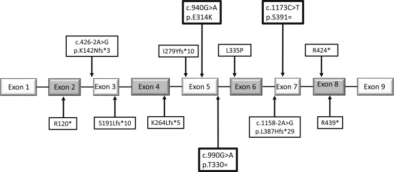
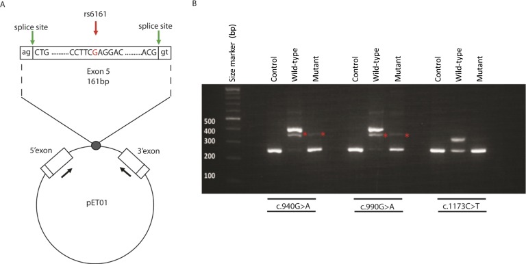
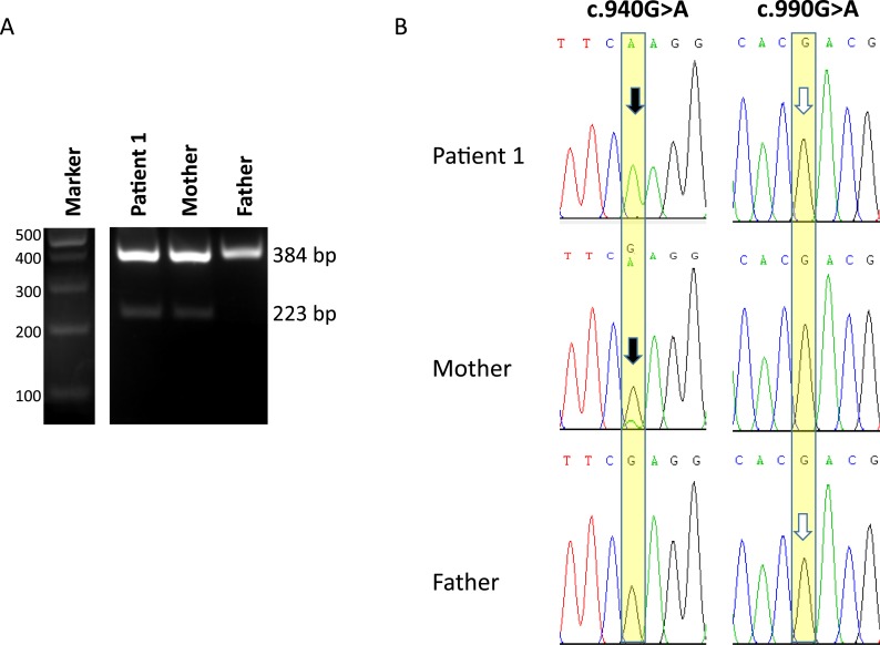
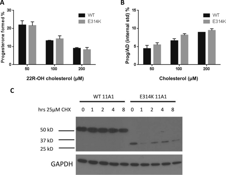
Primary adrenal insufficiency (PAI) is a potentially life-threatening condition that can present with nonspecific features and can be difficult to diagnose. We undertook next generation sequencing in a cohort of children and young adults with PAI of unknown etiology from around the world and identified a heterozygous missense variant (rs6161, c.940G>A, p.Glu314Lys) in CYP11A1 in 19 individuals from 13 different families (allele frequency within undiagnosed PAI in our cohort, 0.102 vs 0.0026 in the Genome Aggregation Database; P < 0.0001). Seventeen individuals harbored a second heterozygous rare disruptive variant in CYP11A1 and two had very rare synonymous changes in trans (c.990G>A, Thr330 = ; c.1173C>T, Ser391 =). Although p.Glu314Lys is predicted to be benign and showed no loss-of-function in an Escherichia coli assay system, in silico and in vitro studies revealed that the rs6161/c.940G>A variant, plus the c.990G>A and c.1173C>T changes, affected splicing and that p.Glu314Lys produces a nonfunctional protein in mammalian cells. Taken together, these findings show that compound heterozygosity involving a relatively common and predicted "benign" variant in CYP11A1 is a major contributor to PAI of unknown etiology, especially in European populations. These observations have implications for personalized management and demonstrate how variants that might be overlooked in standard analyses can be pathogenic when combined with other very rare disruptive changes.

Keywords: Addison disease; CYP11A1; cytochrome p450scc; side chain cleavage enzyme; silent variant.


Figures

Figure 1.
Figure 1.
Position of variants in CYP11A1 genomic/pre-mRNA sequence found in this series of patients with PAI. Boxed in bold, the three predicted benign or synonymous variants assessed for their effect on splicing; SNP rs6161 (c.940G>A, p.Glu314Lys) is 110 bp from the start and 51 bp from the end of exon 5, c.990G>A (p.Thr330 =) occurs at the last base of exon 5, and c.1173C>T (p.Ser391 =) is 16 bp from the start of exon 7.
Figure 2.
Figure 2.
An in vitro assay revealed aberrant splicing of variants c.940G>A, c.990G>A, and c.1173C>T. (A) Minigene construction. Diagram of exon 5 and parts of flanking introns 4 and 5, inserted into the MCS of the pET01 construct (Lower). In the pET01 construct, the intron containing the MCS is flanked by the 5ʹ-donor and 3ʹ-acceptor splice sites of preproinsulin 5ʹ and 3ʹ exons, respectively (green arrows) (http://www.mobitec.com/cms/products/bio/04_vector_sys/exontrap.html). The expression of this vector sequence was driven by the promoter present in the long terminal repeat of Rous sarcoma virus, followed by a short stretch of a eukaryotic gene (phosphatase). The sequences containing the mutations detected in CYP11A1 exons 5 and 7 (mutant) or those that did not (WT) were cloned into the MCS of pET01. The primers used in the RT-PCR experiments within the preproinsulin 5ʹ and 3ʹ exons are indicated by the black arrows (Supplemental Table 5). (B) Representative results of RT-PCR analysis using HEK293 cells transfected with an empty pET01 vector (empty vector), the pET01 vector containing the WT exon or the mutant exon: (Left) the c.940A in exon 5, (Middle) the c.990A change in exon 5, (Right) the C.1174T change in exon 7. A transcript of 225 bp was observed in the empty and mutant vector RT-PCRs, for all variants investigated, corresponding to the two-exon amplification product resulting from splicing of the preproinsulin 5′ and 3′ exons from the vector. For WT vectors containing c.940G and c.990G, a 386 bp transcript was observed corresponding to the three-exon amplification product resulting from correct splicing of CYP11A1 exon 5 between the two vector exons. The intermediate band for exon 5 constructs, at ~350 bp (asterisk), was shown to be a mixture of a sequence that included both 386- and 225-bp bands. For WT c.1173C, a 304-bp transcript corresponding to the size of the three-exon amplification product resulting from correct splicing of CYP11A1 exon 7 was observed. Sanger sequencing confirmed these findings (data not shown).
Figure 3.
Figure 3.
Sequence analysis of in vivo splicing in patient 1 and his parents. (A) RT-PCR amplification products using primers in exons 4 and 6 of CYP11A1. Lanes 1 and 2, patient 1 and his mother, showing two bands corresponding to transcripts containing exon 5 (upper band) and skipping exon 5 (lower band); this was confirmed by Sanger sequencing. In contrast, in lane 3, the father showed only the upper, exon 5 containing, transcript. (B) Partial chromatograms from Sanger sequencing of the upper bands in the patient, mother, and father. For the c.990G>A change (Right), the patient and father’s sequence revealed only the WT c.990G (white arrows), suggesting that the c.990A variant results in exon skipping and destruction of the truncated mRNA by NMD. For c.940G>A (Left), the patient’s sequence only has the mutant c.940A allele A inherited from his mother (black arrow), corroborating the NMD destruction of the c.990A allele inherited from his father; if it were present, it would give a heterozygous base at this position. In contrast, the mother’s sequence shows both WT G and a small peak of the mutant A (black arrow), consistent with skipping of mutant exon 5 in most mutant transcripts but revealing the presence of some transcripts containing exon 5.
Figure 4.
Figure 4.
The p.314Lys protein evaluation. (A) Side-chain cleavage activity of CYP11A1 mutant p.314Lys was unaltered in E. coli. Expression of the purified mutant protein in E. coli showed comparable activity (black bars) as the WT enzyme (gray bars). The activity of the mutant p.314Lys was determined to be indistinguishable from WT protein, whether the substrate was 22R-hydroxycholesterol (Left) or cholesterol (Right). (B) The p.314Lys variant exhibited altered protein expression compared with WT when expressed in HEK-293T cells. HEK-293T cells were transiently transfected with either WT CYP11A1 or p.314Lys constructs and cultured for 48 hours. For the indicated times before protein collection, the cells were treated with 25 µM cycloheximide (CHX). Whole cell lysates were analyzed by immunoblot and probed with anti-CYP11A1 and anti–glyceraldehyde 3-phosphate dehydrogenase (GAPDH) antibodies. For the WT, a full-length protein was observed at 60 kDa. In contrast, for the p.314Lys mutant, the protein was truncated to 30 to 35 kDa, consistent with a cleavage event around the site of the amino acid change. CHX treatment revealed the mutant protein also had a shorter half-life.

References

    1. Miller WL, Auchus RJ. The molecular biology, biochemistry, and physiology of human steroidogenesis and its disorders. Endocr Rev. 2011;32(1):81–151. - PMC - PubMed
    1. Miller WL. Why nobody has P450scc (20,22 desmoslase) deficiency. J Clin Endocrinol Metab. 1998;83(4):1399–1400. - PubMed
    1. Guran T, Buonocore F, Saka N, Ozbek MN, Aycan Z, Bereket A, Bas F, Darcan S, Bideci A, Guven A, Demir K, Akinci A, Buyukinan M, Aydin BK, Turan S, Agladioglu SY, Atay Z, Abali ZY, Tarim O, Catli G, Yuksel B, Akcay T, Yildiz M, Ozen S, Doger E, Demirbilek H, Ucar A, Isik E, Ozhan B, Bolu S, Ozgen IT, Suntharalingham JP, Achermann JC. Rare causes of primary adrenal insufficiency: genetic and clinical characterization of a large nationwide cohort. J Clin Endocrinol Metab. 2016;101(1):284–292. - PMC - PubMed
    1. Tee MK, Abramsohn M, Loewenthal N, Harris M, Siwach S, Kaplinsky A, Markus B, Birk O, Sheffield VC, Parvari R, Hershkovitz E, Miller WL. Varied clinical presentations of seven patients with mutations in CYP11A1 encoding the cholesterol side-chain cleavage enzyme, P450scc. J Clin Endocrinol Metab. 2013;98(2):713–720. - PMC - PubMed
    1. Parajes S, Chan AO, But WM, Rose IT, Taylor AE, Dhir V, Arlt W, Krone N. Delayed diagnosis of adrenal insufficiency in a patient with severe penoscrotal hypospadias due to two novel P450 side-change cleavage enzyme (CYP11A1) mutations (p.R360W; p.R405X). Eur J Endocrinol. 2012;167(6):881–885. - PMC - PubMed

LinkOut - more resources