

Inhibition of Ephrin-B2 in brain pericytes decreases cerebral pathological neovascularization in diabetic rats
- PMID: 30620753
- PMCID: PMC6324788
- DOI: 10.1371/journal.pone.0210523
Inhibition of Ephrin-B2 in brain pericytes decreases cerebral pathological neovascularization in diabetic rats
Abstract
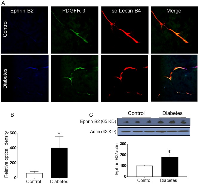
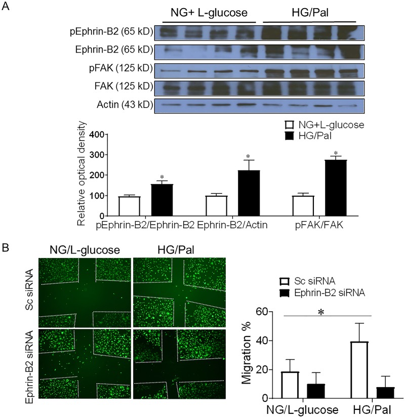
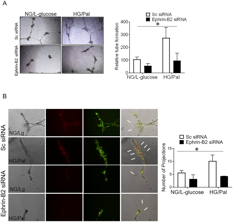
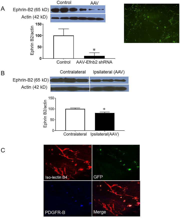
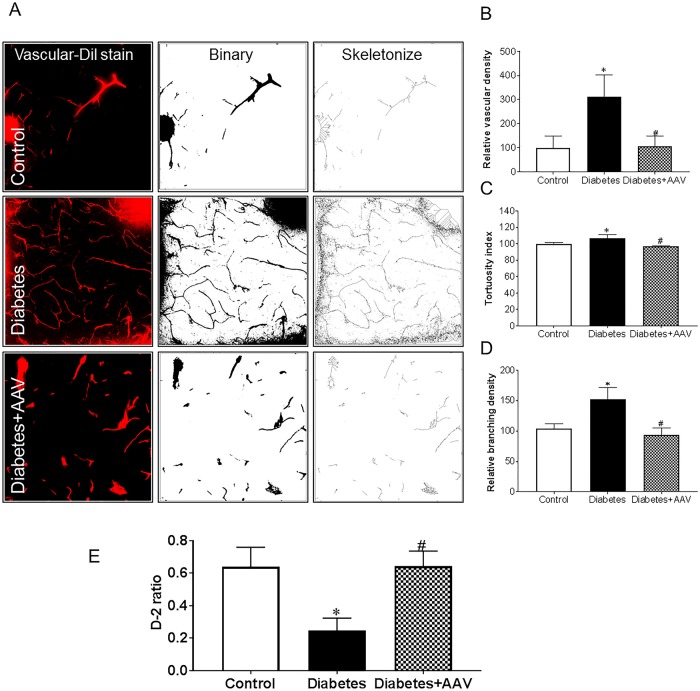
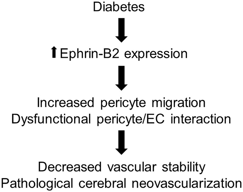
We have previously shown that diabetes causes dysfunctional cerebral neovascularization that increases the risk for cerebrovascular disorders such as stroke and cognitive impairment. Pericytes (PCs) play a pivotal role in the angiogenic process through their interaction with the endothelial cells (EC). Yet, the role of PCs in dysfunctional cerebral neovascularization in diabetes is unclear. In the present study, we tested the hypothesis that the increased proangiogenic Ephrin-B2 signaling in PCs contributes to the dysfunctional cerebral neovascularization in diabetes. Type-II diabetes was induced by a combination of high fat diet and low dose streptozotocin injection in male Wistar rats. Selective in vivo Ephrin-B2 silencing in brain PCs was achieved using the stereotactic injection of adeno-associated virus (AAV) with NG2-promoter that expresses Ephrin-B2 shRNA. Neovascularization was assessed using vascular fluorescent dye stain. Novel object recognition (NOR) test was used to determine cognitive functions. Human brain microvascular pericytes HBMVPCs were grown in high glucose 25 mM and palmitate 200 uM (HG/Pal) to mimic diabetic conditions. Scratch migration and tube formation assays were conducted to evaluate PC/EC interaction and angiogenic functions in PC/EC co-culture. Diabetes increased the expression of Ephrin-B2 in the cerebrovasculature and pericytes. Concomitant increases in cerebral neovascularization parameters including vascular density, tortuosity and branching density in diabetic rats were accompanied by deterioration of cognitive function. Inhibition of Ephrin-B2 expression in PCs significantly restored cerebral vascularization and improved cognitive functions. HG/Pal increased PC/EC angiogenic properties in co-culture. Silencing Ephrin-B2 in PCs significantly reduced PC migration and PC/EC co-culture angiogenic properties. This study emphasizes the significant contribution of PCs to the pathological neovascularization in diabetes. Our findings introduce Ephrin-B2 signaling as a promising therapeutic target to improve cerebrovascular integrity in diabetes.
Conflict of interest statement
The authors declare that there is no duality of interest associated with this manuscript.
Figures
Similar articles
-
Increased Ephrin-B2 expression in pericytes contributes to retinal vascular death in rodents.Vascul Pharmacol. 2020 Aug;131:106761. doi: 10.1016/j.vph.2020.106761. Epub 2020 Jun 22. Vascul Pharmacol. 2020. PMID: 32585189 Free PMC article.
-
GLP-1 receptor nitration contributes to loss of brain pericyte function in a mouse model of diabetes.Diabetologia. 2022 Sep;65(9):1541-1554. doi: 10.1007/s00125-022-05730-5. Epub 2022 Jun 10. Diabetologia. 2022. PMID: 35687178 Free PMC article.
-
Platelet-derived growth factor signaling through ephrin-b2 regulates hepatic vascular structure and function.Gastroenterology. 2008 Aug;135(2):671-9. doi: 10.1053/j.gastro.2008.04.010. Epub 2008 Apr 16. Gastroenterology. 2008. PMID: 18570897 Free PMC article.
-
Regulation of signaling interactions and receptor endocytosis in growing blood vessels.Cell Adh Migr. 2014;8(4):366-77. doi: 10.4161/19336918.2014.970010. Cell Adh Migr. 2014. PMID: 25482636 Free PMC article. Review.
-
The role of endothelial cell-pericyte interactions in vascularization and diseases.J Adv Res. 2025 Jan;67:269-288. doi: 10.1016/j.jare.2024.01.016. Epub 2024 Jan 20. J Adv Res. 2025. PMID: 38246244 Free PMC article. Review.
Cited by
-
Not All Lectins Are Equally Suitable for Labeling Rodent Vasculature.Int J Mol Sci. 2021 Oct 26;22(21):11554. doi: 10.3390/ijms222111554. Int J Mol Sci. 2021. PMID: 34768985 Free PMC article.
-
Increased Ephrin-B2 expression in pericytes contributes to retinal vascular death in rodents.Vascul Pharmacol. 2020 Aug;131:106761. doi: 10.1016/j.vph.2020.106761. Epub 2020 Jun 22. Vascul Pharmacol. 2020. PMID: 32585189 Free PMC article.
-
GLP-1 receptor nitration contributes to loss of brain pericyte function in a mouse model of diabetes.Diabetologia. 2022 Sep;65(9):1541-1554. doi: 10.1007/s00125-022-05730-5. Epub 2022 Jun 10. Diabetologia. 2022. PMID: 35687178 Free PMC article.
-
Immunomodulatory Approaches in Diabetes-Induced Cardiorenal Syndromes.Front Cardiovasc Med. 2021 Jan 28;7:630917. doi: 10.3389/fcvm.2020.630917. eCollection 2020. Front Cardiovasc Med. 2021. PMID: 33585587 Free PMC article. Review.
-
Platelet-derived growth factor signaling in pericytes promotes hypothalamic inflammation and obesity.Mol Med. 2024 Feb 5;30(1):21. doi: 10.1186/s10020-024-00793-z. Mol Med. 2024. PMID: 38317079 Free PMC article.
References
-
- Herman WH, Dasbach EJ, Songer TJ, Eastman RC. The cost-effectiveness of intensive therapy for diabetes mellitus. Endocrinol Metab Clin North Am. 1997;26(3):679–95. . - PubMed
Publication types
MeSH terms
Substances
Grants and funding
LinkOut - more resources
Full Text Sources
Medical
