. 2019 Jan 8;14(1):e0210474.
doi: 10.1371/journal.pone.0210474. eCollection 2019.

Formation of high molecular weight p62 by CORM-3

Affiliations

Formation of high molecular weight p62 by CORM-3

Toshihiko Aki et al. PLoS One. .

Abstract

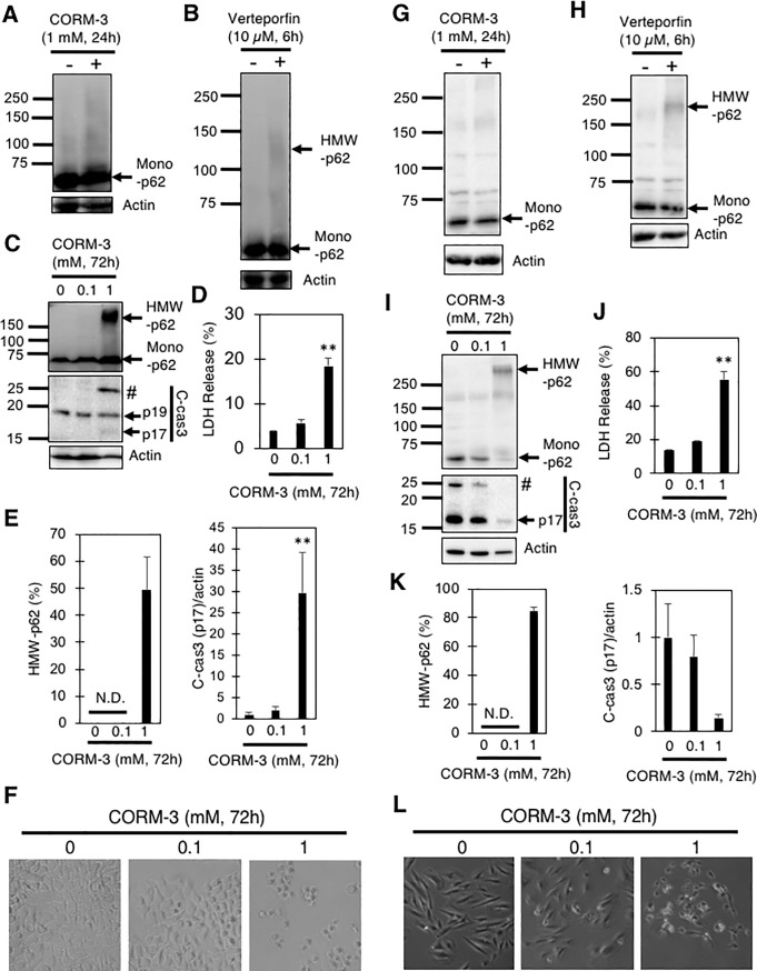
CORM-3 is a water-soluble carbon monoxide (CO)-releasing molecule developed for possible therapeutic use of CO. CORM-3 belongs to a group of metal carbonyl compounds that contain transition metals and carbonyls as the central scaffold and coordinated ligands, respectively. CORM-3 has been reported to be reactive with many proteins in eukaryotes including mammals. Among them, several extracellular proteins, such as lysozyme, as well as plasma albumin and fibronectin, have been shown to interact directly with CORM-3. p62 is an intracellular adaptor protein required for targeting ubiquitinated (Ub) proteins to lysosomal degradation through autophagy. p62 has been shown to undergo self-oligomerization via covalent crosslinking in response to treatment with verteporfin, a benzoporphyrin derivative used for photodynamic therapy. Here we show that CORM-3 also interacts directly with p62. When applied to mouse embryonic fibroblasts (MEFs) at a high concentration (1 mM), CORM-3 causes the formation of reduction- and detergent-resistant high molecular weight (HMW)-p62. HMW-p62 accumulates more in atg5-/- MEFs than in wild type (WT) MEFs, showing the elimination of HMW-p62 through autophagy. HMW-p62 is also generated in H9c2 rat cardiomyoblastoma as well as A549 human alveolar epithelial cells, suggesting that HMW-p62 formation is not specific to MEFs, but, rather, is a general event in mammalian cells. HMW-p62 formation by CORM-3 can be reproduced using purified p62 in vitro, demonstrating the direct interaction between CORM-3 and p62. These results show that p62 is a CORM-3-interactive intracellular protein.


Conflict of interest statement

The authors have declared that no competing interests exist.

Figures

Fig 1
Fig 1. CORM-3 induces the death of MEFs.
(A and B) MEFs were treated with the indicated concentrations (0, 0.1, or 1 mM) of CORM-3 for 24 or 48 hours (A), and the relative levels of p17 fragment of cleaved-caspase-3 to actin (B and C) as well as LDH release into the medium (D and E) are shown. #, uncharacterized fragment. Data represent the means and S.E. *, p<0.05; **, p<0.01 by Turkey-Kramer’s test (B and E, n = 4). *, p<0.05; **, p<0.01 by Bonferroni’s test (C, n = 3).
Fig 2
Fig 2. Generation of HMW-p62 by CORM-3 in MEFs.
(A and B) MEFs treated with 0, 0.1, or 1 mM CORM-3 for 24 hours were subjected to immunoblot analysis using anti-LC3 and -p62 antibodies. Relative levels of LC3-II, as well as HMW- and mono-p62, to actin are shown as means and S.E. (n = 4). **, p<0.01 versus 0 mM by Dunnett’s test. (C) p62 dot formation in WT and atg5-/- MEFs treated with or without 1 mM CORM-3 for 24 hours. Immunofluorescence analysis was performed using anti-p62 antibody and Alexa488 (green)-labeled anti-IgG antibody. (D) Atg5-/-MEFs were treated with or without 1 mM CORM-3 for 30 hours and cell lysates were subjected to SDS-PAGE with or without the reduction by 2-mercaptoethanol (2-ME). Immunoblot analysis were performed using anti-p62 antibody.
Fig 3
Fig 3. Generation of HMW-p62 by verteporfin in MEFs.
(A) MEFs treated with 10 μM verteporfin for 6 hours were subjected to immunoblot analysis using anti-p62 antibody. Relative levels of p62 to actin are shown as means and S.E. (n = 4). (B) Lack of HMW-ROCK-1 in WT and atg5-/-MEFs treated with verteporfin or CORM-3. MEFs treated with 10 μM verteporfin for 6 hours or 1 mM CORM-3 for 24 hours were subjected to immunoblot analysis using anti-ROCK-1 antibody.
Fig 4
Fig 4. Generation of HMW-FN by CORM-3 in A549 and H9c2 cells.
A549 cells (A-E) or H9c2 cells (G-K) treated with verteporfin (10 μM, 6 hours) or CORM-3 (0–1 mM, 24 or 72 hours) were subjected to immunoblot analysis using anti-p62 and anti-cleaved caspase 3 (c-cas3) antibodies. #, uncharacterized fragment. Percentages of HMW-p62 in total (HMW+mono) p62 and relative levels of c-cas3 (p17) to actin are also shown. (D and J) Percentages of LDH released from A549 (D) and H9c2 (J) cells into the medium 72 hours after treatment with the indicated concentrations of CORM-3. Graphs show means and S.E. (n = 4). **, p<0.01 versus control by Dunnett’s test. N.D., not detected. Representative phase contrast images of A549 (F) and H9c2 (L) cells treated with or without CORM-3 at the indicated concentrations for 72 hours are also shown.
Fig 5
Fig 5. Production of HMW-p62 by CORM-3 in vitro.
Purified recombinant full-length human p62 (A), bovine plasma fibronectin (FN) (B), or bovine serum albumin (BSA) (C) was dissolved in PBS and treated with CORM-3 (1 mM), CORM-A1 (1 mM), or RuCl3 (1 mM) for 0–60 min. p62, FN, and BSA were also treated with verteporfin (10 μM) for 0-60min. After SDS-PAGE, p62 and FN were visualized by immunoblot analysis while BSA was stained with CBB.
Fig 6
Fig 6. Co-localization of p62 dots with Ub-positive aggregates, but not with LC3-positive puncta, in atg5-/- MEFs.
WT (A) and atg5-/- (B) MEFs were treated with or without 1 mM CORM-3 for 24 hours, and immunocytochemistry was performed using anti-ubiquitin conjugated protein (Ub), anti-p62, and anti-LC3 antibodies. Alexa488 (green)- and Alexa549 (red)-conjugated anti-IgG antibodies were used as secondary antibodies to visualize antigens under a fluorescence microscope. White and yellow arrows indicate Ub- and LC3-positive structures, respectively. Percentages of LC3-positive/Ub-negative (LC3+/Ub-), LC3-positive/Ub-positive (LC3+/Ub+), and LC3-negative/Ub-positive (LC3-/Ub+) dots, as well as p62-positive/Ub-negative (p62+/Ub-), p62-positive/Ub-positive (p62+/Ub+), and p62-negative/Ub-positive (p62-/Ub+) dots, in atg5-/- MEFs treated with 1 mM CORM-3 were shown (C).
Fig 7
Fig 7. Effects of siRNA-mediated knock-down of p62 on CORM-3-induced death of atg5-/- MEFs.
(A) WT and atg5-/- MEFs were treated with a control siRNA (C) or a siRNA for p62 (p62) and examined for their intracellular levels of p62 (left panels) by immunoblotting. #, uncharacterized fragment. The graphs show the means and S.E. of four samples. **, p<0.01 by student’s t-test. (B) SiRNA-treated MEFs were also exposed to 1 mM CORM-3 for 48 hours, and the relative levels of p17 fragment of cleaved-caspase-3 (c-cas3) were determined by immunoblotting. The graphs show the means and S.E. of four samples. *, p<0.05; **, p<0.01 by Turkey-Kramer’s test. Representative images of atg5-/- MEFs treated with or without the siRNA for p62 and then incubated with 1 mM CORM-3 for 48 hours, are shown (C). Percentages of LDH into the medium is also shown (D).

References

    1. Clark JE, Naughton P, Shurey S, Green CJ, Johnson TR, Mann BE, et al. Cardioprotective actions by a water-soluble carbon monoxide-releasing molecule. Circ Res. 2003;93 (2): e2–8 10.1161/01.RES.0000084381.86567.08 - DOI - PubMed
    1. Motterlini R, and Otterbein LE. The therapeutic potential of carbon monoxide. Nat Rev Drug Discov. 2010;9 (9): 728–743 10.1038/nrd3228 - DOI - PubMed
    1. Jung SS, Moon JS, Xu JF, Ifedigbo E, Ryter SW, Choi AM, et al. Carbon monoxide negatively regulates NLRP3 inflammasome activation in macrophages. Am J Physiol Lung Cell Mol Physiol. 2015;308 (10): L1058–1067 10.1152/ajplung.00400.2014 - DOI - PMC - PubMed
    1. Hoetzel A, Dolinay T, Schmidt R, Choi AM, and Ryter SW. Carbon monoxide in sepsis. Antioxid Redox Signal. 2007;9 (11): 2013–2026 10.1089/ars.2007.1762 - DOI - PubMed
    1. Nakahira K, and Choi AM. Carbon monoxide in the treatment of sepsis. Am J Physiol Lung Cell Mol Physiol. 2015;309 (12): L1387–1393 10.1152/ajplung.00311.2015 - DOI - PMC - PubMed

Publication types

MeSH terms