Review
. 2019 Jan 6;24(1):186.
doi: 10.3390/molecules24010186.

Vibrational Approach to the Dynamics and Structure of Protein Amyloids

Affiliations
Review

Vibrational Approach to the Dynamics and Structure of Protein Amyloids

Haoqian Li et al. Molecules. .

Abstract

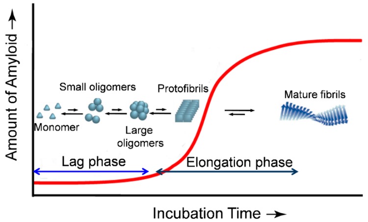
Amyloid diseases, including neurodegenerative diseases such as Alzheimer's and Parkinson's, are linked to a poorly understood progression of protein misfolding and aggregation events that culminate in tissue-selective deposition and human pathology. Elucidation of the mechanistic details of protein aggregation and the structural features of the aggregates is critical for a comprehensive understanding of the mechanisms of protein oligomerization and fibrillization. Vibrational spectroscopies, such as Fourier transform infrared (FTIR) and Raman, are powerful tools that are sensitive to the secondary structure of proteins and have been widely used to investigate protein misfolding and aggregation. We address the application of the vibrational approaches in recent studies of conformational dynamics and structural characteristics of protein oligomers and amyloid fibrils. In particular, introduction of isotope labelled carbonyl into a peptide backbone, and incorporation of the extrinsic unnatural amino acids with vibrational moieties on the side chain, have greatly expanded the ability of vibrational spectroscopy to obtain site-specific structural and dynamic information. The applications of these methods in recent studies of protein aggregation are also reviewed.

Keywords: Raman; amyloid; infrared; isotopic labelling; oligomers; protein aggregation; site-specific probe; vibrational spectroscopy.


Conflict of interest statement

The authors state that they have no conflicts of interest.

Figures

Figure 1
Figure 1
Schematic representation of a typical nucleated polymerization process of protein aggregation. Oligomeric nuclei are formed in the early lag phase stage, being a critical rate limiting step. In an elongation phase, addition of monomers and/or oligomers onto the nucleus allows formation of fibrils which is energetically favorable.
Figure 2
Figure 2
Left panel: normalized FTIR spectra of Aβ1–40 monomers (M) in 10 mM HEPES/D2O pD11, fibrils (F) after 24 h incubation in 10 mM HEPES pD7.4, and Cu(II)-induced oligomers (Aβ1–40-Cu(II)Õ) after 24 h incubation at 37 °C, pD7.4 [51]. Right panel: second derivatives of the FTIR spectra. Reprinted with permission.
Figure 3
Figure 3
2D-IR spectra and kinetics curves of human IAPP with 13C=18O labeled at the Ala25 residue [113]. (A) The first 2D-IR spectrum at t = 5 min. (BD) Difference 2D-IR spectra at t = 31, 66, and 205 min, calculated by subtracting the t = 5 min spectrum. Black boxes surround the spectral features of the unlabeled portion of the peptide, whereas red boxes enclose the diagonal peaks of the isotope labeled Ala25. Blue and green arrows highlight the 2 labeled features, whereas magenta and red arrows point to the cross-peak between the 13C=18O Ala25 and the unlabeled β-sheet. (E) Kinetics of the diagonal peaks of the unlabeled β-sheet at 1617 cm−1 and the 2 label features (blue and green arrows). (F) Comparison of the kinetics of the cross-peak and the diagonal peaks. Reprinted with permission.
Figure 4
Figure 4
Raman spectra of three Aβ1–23 mutants before and after being incubated for 3 day for aggregation [172]. In the mutants, the PheCN residue was used to replace Tyr10 (Aβ1–23M1), Phe19 (Aβ1–23M2), and Phe20 (Aβ1–23M3), respectively. The vertical dashed lines indicate Raman wavenumbers at 2229 and 2237 cm−1, respectively. Reprinted with permission.

References

    1. Selkoe D.J. Folding proteins in fatal ways. Nature. 2003;426:900–904. doi: 10.1038/nature02264. - DOI - PubMed
    1. Chiti F., Dobson C.M. Protein misfolding, functional amyloid, and human disease. Annu. Rev. Biochem. 2006;75:333–366. doi: 10.1146/annurev.biochem.75.101304.123901. - DOI - PubMed
    1. Sipe J.D., Cohen A.S. Review: History of the amyloid fibril. J. Struct. Biol. 2000;130:88–98. doi: 10.1006/jsbi.2000.4221. - DOI - PubMed
    1. Jahn T.R., Makin O.S., Morris K.L., Marshall K.E., Tian P., Sikorski P., Serpell L.C. The common architecture of cross-β amyloid. J. Mol. Biol. 2010;395:717–727. doi: 10.1016/j.jmb.2009.09.039. - DOI - PubMed
    1. Eisenberg D., Jucker M. The amyloid state of proteins in human diseases. Cell. 2012;148:1188–1203. doi: 10.1016/j.cell.2012.02.022. - DOI - PMC - PubMed

LinkOut - more resources