. 2019 Jan 6;20(1):180.
doi: 10.3390/ijms20010180.

Ontogenetic Changes in Auxin Biosynthesis and Distribution Determine the Organogenic Activity of the Shoot Apical Meristem in pin1 Mutants

Affiliations

Ontogenetic Changes in Auxin Biosynthesis and Distribution Determine the Organogenic Activity of the Shoot Apical Meristem in pin1 Mutants

Alicja Banasiak et al. Int J Mol Sci. .

Abstract

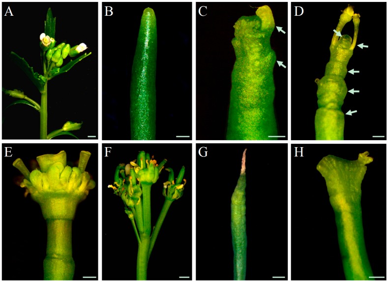
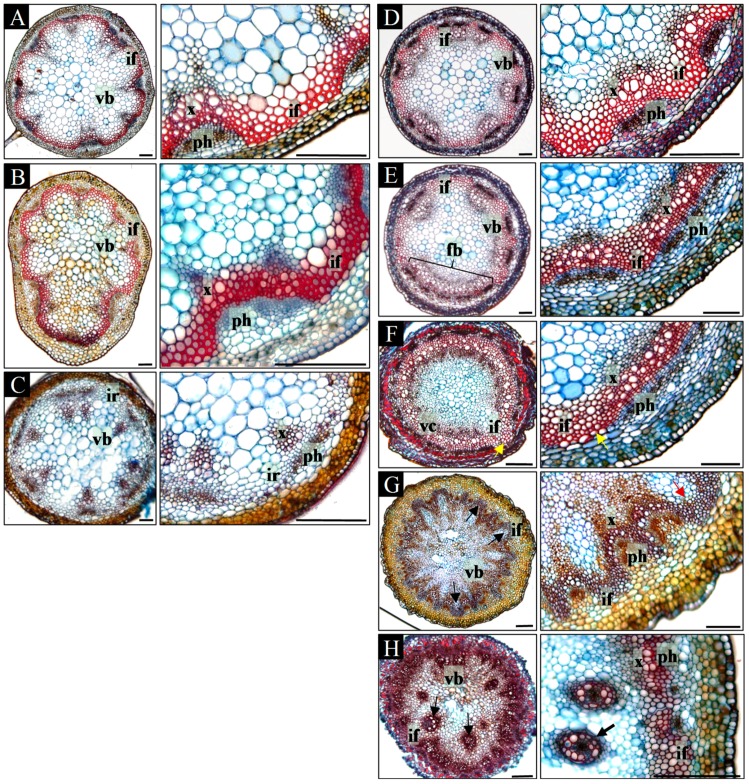
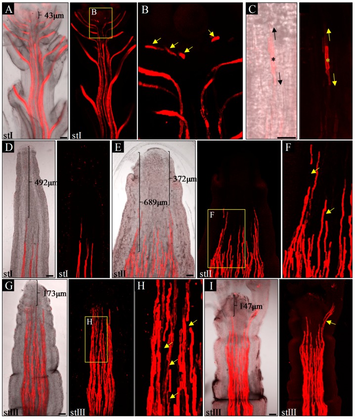
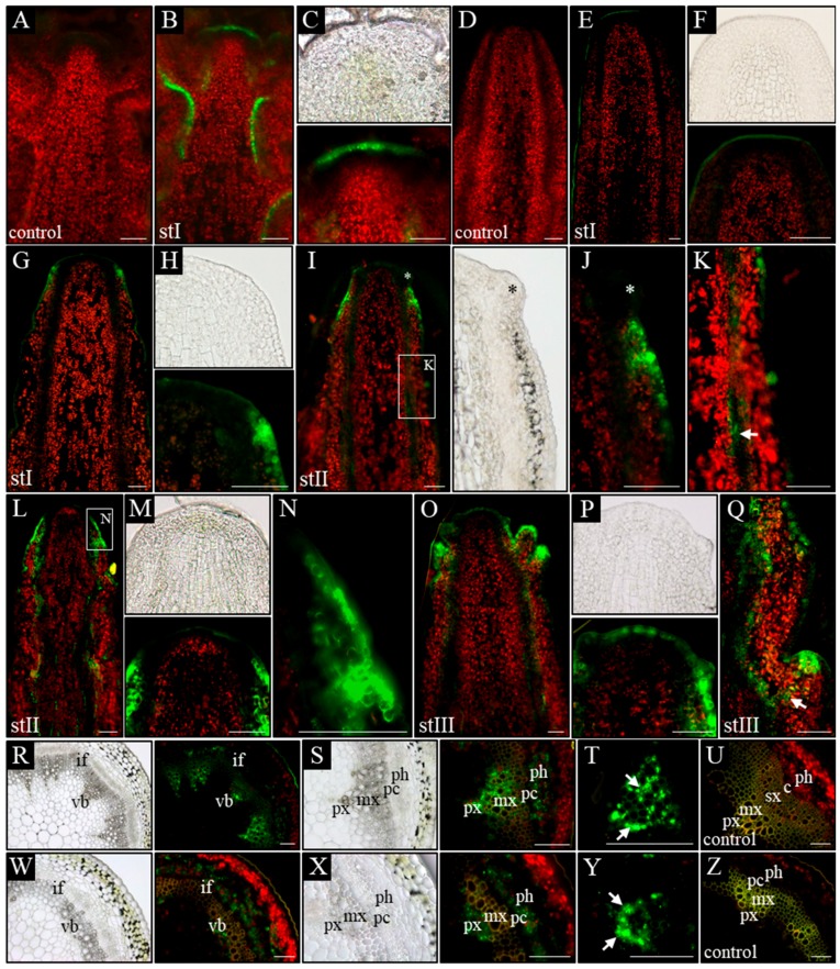
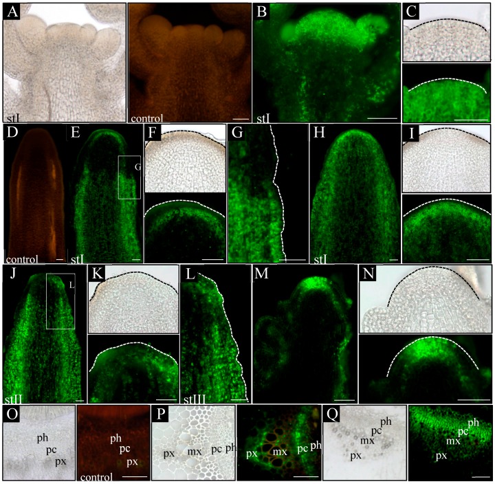
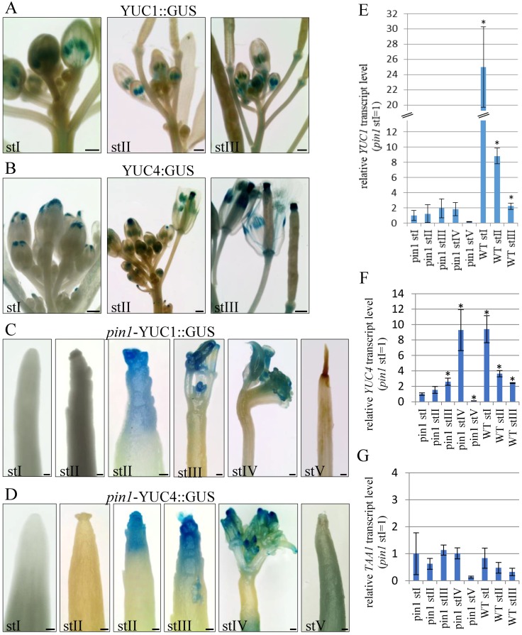
In the shoot apical meristem (SAM) of Arabidopsis, PIN1-dependent polar auxin transport (PAT) regulates two crucial developmental processes: organogenesis and vascular system formation. However, the knockout mutation in the PIN1 gene does not fully inhibit these two processes. Therefore, we investigated a potential source of auxin for organogenesis and vascularization during inflorescence stem development. We analyzed auxin distribution in wild-type (WT) and pin1 mutant plants using a refined protocol of auxin immunolocalization; auxin activity, with the response reporter pDR5:GFP; and expression of auxin biosynthesis genes YUC1 and YUC4. Our results revealed that regardless of the functionality of PIN1-mediated PAT, auxin is present in the SAM and vascular strands. In WT plants, auxin always accumulates in all cells of the SAM, whereas in pin1 mutants, its localization within the SAM changes ontogenetically and is related to changes in the structure of the vascular system, organogenic activity of SAM, and expression levels of YUC1 and YUC4 genes. Our findings indicate that the presence of auxin in the meristem of pin1 mutants is an outcome of at least two PIN1-independent mechanisms: acropetal auxin transport from differentiated tissues with the use of vascular strands and auxin biosynthesis within the SAM.

Keywords: Arabidopsis; PAT; SAM; YUC genes; auxin immunolocalization; organogenesis; pin1 mutant; vascular system; xylem.


Conflict of interest statement

The authors declare no conflict of interest.

Figures

Figure 1
Figure 1
The phenotype of inflorescence shoots. The phenotype of the stem apex of wild-type (WT) (A) and pin1 mutant (BH) Arabidopsis plants. pin1 mutant stems showing several developmental features of its apex: without organs (B); with single bulges and developing organ primordia (C), denoted with arrows; with numerous bulges, folds and malformed organs (D), denoted with arrows; a termination with a single organ (E), multiple organs (F), and meristem necrosis (G); fasciation (H). Scale bar 200 μm.
Figure 2
Figure 2
Ontogenetic changes in the structure of the inflorescence shoots. Subsequent cross-sections of the inflorescence shoot of WT (AC) and pin1 mutant (DH) plants, showed in the basal-apical sequence. The left picture presents the vascular strands arrangement, and the right picture the magnification of the interfascicular region. The stem of the WT plant has in its basal part discrete vascular bundles (A); under the first branch increased number of bundles (B); and in the apical region the stem lacks differentiated interfascicular fibers (C). The stem of the pin1 mutant is characterized by (looking from the most basal part of the stem towards the top) regions with: discrete vascular bundles (D); fusing bundles (E), denoted with bracket; the vascular cylinder, with interfascicular fibers displaced towards the pith (F), direction of fibers displacement denoted with arrow; vascular cylinder splitting into numerous small bundles, with interfascicular fibers displaced towards the stem surface (G), direction of fibers displacement denoted with arrows; concentric bundles in the pith of the stem (H), denoted with arrows. vb—vascular bundle, fv—fusing vascular bundles, vc—vascular cylinder, if—interfascicular fibers, ir—interfascicular region, x—xylem, ph—phloem. Scale bar 100 μm.
Figure 3
Figure 3
Structure of the xylem strands in the apices of the inflorescence stems, stained with propidium iodide. In WT plants, xylem strands differentiate close to the meristem—distance denoted with bracket, and are continuous in the mature stem and organs (A); discontinuities (denoted with arrows) are present only in the apical, meristematic region of the shoot (B). The discontinuities in the xylem strands, however, disappear during development, as a result of bidirectional xylem differentiation—denoted with arrows (C). pin1 mutants are characterized by: discrete, continuous xylem strands differentiating far from the meristem (distance denoted with brackets), in stage I (D); increased number of xylem strands, in comparison to stage I, with variable distance from the meristem—denoted with brackets (E) and with local discontinuities—denoted with arrows (F), in stage II; numerous differentiated xylem strands close to the meristem—distance denoted with bracket (G), with frequent discontinuities—denoted with arrows (H), and developing malformed organs with their own xylem strands connected to the strands of the main stem—denoted by arrow (I), in stage III. St I–III—successive developmental stages, the asterisk—first differentiated protoxylem element. Scale bar 50 μm.
Figure 4
Figure 4
Spatial localization of the auxin response reporter pDR5:GFP activity. Longitudinal (AQ) and transversal (RZ) sections through the apical part of inflorescence stems of WT (AC,RU) and pin1 mutant (DQ,WZ) plants. On the longitudinal sections, in WT plants, the GFP signal is visible in the vascular strands (B) and the L1 layer of the meristem (C). In the pin1 mutant, in stage I, the activity of the pDR5:GFP transgene is not detected (E,F) or is present in the L1–L2 layers of the peripheral zone of the meristem (G,H); in stage II, GFP signal accumulates in the surface layers at the base of the organ primordia—primordium denoted with asterisk (I,J) and locally in the vascular bundles - denoted with arrow (K) or in the L1–L2 layers of the peripheral zone of the SAM and in all vascular bundles (LN); in stage III, the GFP signal is visible in the L1 layer of the entire meristem, at the tip of the developing organs and in all vascular strands (O,P) as well as of the cells (denoted with arrow) connecting the tip of an organ primordia with the vasculature (Q). On the transversal sections, the activity of the pDR5:GFP transgene in WT plants is visible in the phloem, xylem and locally in procambial cells (RU). In terms of the xylem, GFP signal accumulates in all xylem parenchyma cells—denoted with arrows (S,T). In the pin1 mutant GFP signal is visible in xylem and phloem (WZ). In terms of the xylem, GFP signal is present only in the parenchyma cells on the protoxylem side—denoted with arrows (Y). (A,D,U,Z) Tissue autofluorescence (control); (C,F,H,J,M,P) magnifications of the meristems. St I–III—successive developmental stages; vb—vascular bundle, if—interfascicular fibers, px—protoxylem, mx—metaxylem, sx—secondary xylem, ph—phloem, pc-procambium, c—cambium. Scale bare 50 μm.
Figure 5
Figure 5
Immunolocalization of auxin in the inflorescence stems. Longitudinal (AN) and transversal (OQ) sections through the apical part of the inflorescence stems of WT (AC,OP) and pin1 mutant (DN,Q) plants. On the longitudinal sections, in WT plants, the strongest signal of auxin immunolocalization is visible in the entire meristem, developing organs and all vascular strands (B,C). In the pin1 mutant, in stage I, the auxin signal is visible in the L3 layer of the meristem, vascular strands of the apical part of stem (E,F) and in all cell types of the more basal stem region (G), or in all cells of the entire shoot, except the surface layers (H,I); in stage II, the auxin immunolocalization signal is additionally visible in the L1 layer of the peripheral zone (J,K), and the more basal region, characterized by the presence of the auxin signal in all cells, is closer to the meristem (L); in stage III, the immunolocalization signal is visible in the L1–L3 layers of the entire meristem, the surface layer of the organs and all vascular strands (M,N). On the transversal sections, in WT plants, auxin immunolocalization signal accumulates in the procambial region, phloem and all cells of xylem parenchyma (P). In the pin1 mutant, the signal is visible in the procambial region, phloem, and xylem parenchymal cells on the protoxylem side (Q). (A,D,O) Tissue autofluorescence (control); (C,F,I,K,N) magnifications of the meristems, meristem outlined with dashed line; (G,L) magnifications of the marked regions, the surface of stem outlined with dashed line. St I–III—successive developmental stages; px—protoxylem; mx—metaxylem; ph—phloem; pc—procambium. Scale bar 50 μm.
Figure 6
Figure 6
Expression analyses of auxin biosynthesis genes YUC1 and YUC4. (AD) Activity of the YUC1 (A,C) and YUC4 (B,D) promoters visualized by the activity of the GUS reporter protein (blue). In WT plants, in stages I-III, activity of the pYUC1:GUS (A) and pYUC4:GUS (B) reporters is visible in developing organs. In pin1 mutants the expression of pYUC1:GUS (C) and pYUC4:GUS (D) is not detected in stage I plants; is visible in some shoots with organ primordia in stage II plants; is present in most plants from stages III and IV; and is not detected in stage V plants. (EG) The comparison of YUC1 (E), YUC4 (F) and TAA1 (G) transcript levels during different ontogenetic stages of inflorescence shoots development of WT and pin1 mutant plants. Abundance of the transcripts, in each case, is relative to the pin1 stage I sample. The level of YUC1 and YUC4 genes expression in pin1 mutants increases during ontogenesis, but yet is generally weaker in comparison to WT plants. The level of TAA1 gene expression in WT plants and pin1 mutants does not generally significantly differ. Statistically significant differences to the pin1 stage I sample are denoted with asterisk (t-test, significance at p = 0.05). St. I–V (pin 1) and St. I–III (WT)—successive developmental stages. Scale bar 500 μm.

References

    1. Reinhardt D., Pesce E.R., Stieger P., Mandel T., Baltensperger K., Bennett M., Traas J., Friml J., Kuhlemeier C. Regulation of phyllotaxis by polar auxin transport. Nature. 2003;426:255–260. doi: 10.1038/nature02081. - DOI - PubMed
    1. Dengler N.G. The shoot apical meristem and development of vascular architecture. Can. J. Bot. 2006;84:1660–1671. doi: 10.1139/b06-126. - DOI
    1. Steeves T.A., Sussex I.A. Patterns in Plant Development. Cambridge University Press; Cambridge, UK: 1989.
    1. Reddy G.V., Meyerowitz E.M. Stem-cell homeostasis and growth dynamics can be uncoupled in the Arabidopsis shoot apex. Science. 2005;310:663–667. doi: 10.1126/science.1116261. - DOI - PubMed
    1. Rieu I., Laux T. Signaling pathways maintaining stem cells at the plant shoot apex. Semin. Cell Dev. Biol. 2009;20:1083–1088. doi: 10.1016/j.semcdb.2009.09.013. - DOI - PubMed

MeSH terms

LinkOut - more resources