

Clonorchiasis sinensis detected by laparoscopic exploration of biliary tracts in two patients with obstructive jaundice
- PMID: 30621611
- PMCID: PMC6325787
- DOI: 10.1186/s12879-019-3679-y
Clonorchiasis sinensis detected by laparoscopic exploration of biliary tracts in two patients with obstructive jaundice
Abstract
Background: Hepatic clonorchiasis is one of the most prevalent food-borne parasitic diseases worldwide. Clonorchis sinensis, the pathogen, is the major parasitic trigger contributing to cholangitis, cholelithiasis, and even cholangiocarcinoma. Unfortunately, unspecific clinical manifestations of patients with hepatic clonorchiasis tend to mislead clinicians to neglect or misdiagnose them, following ignorance of appropriate therapy. Our case report may shed light on definite diagnosis of clonorchiasis with concomitant cholelithiasis, methodology for surgical drainage of the parasites, and postoperative anthelmintic therapy.
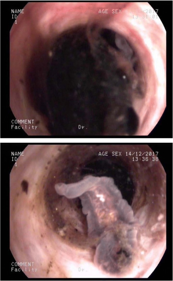
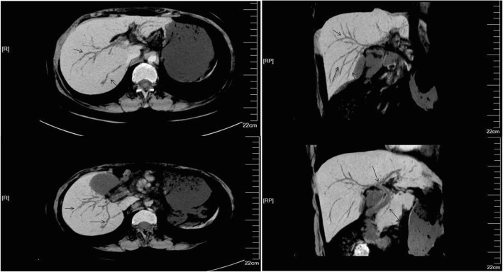
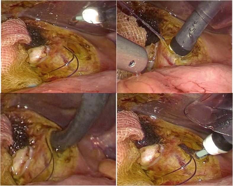
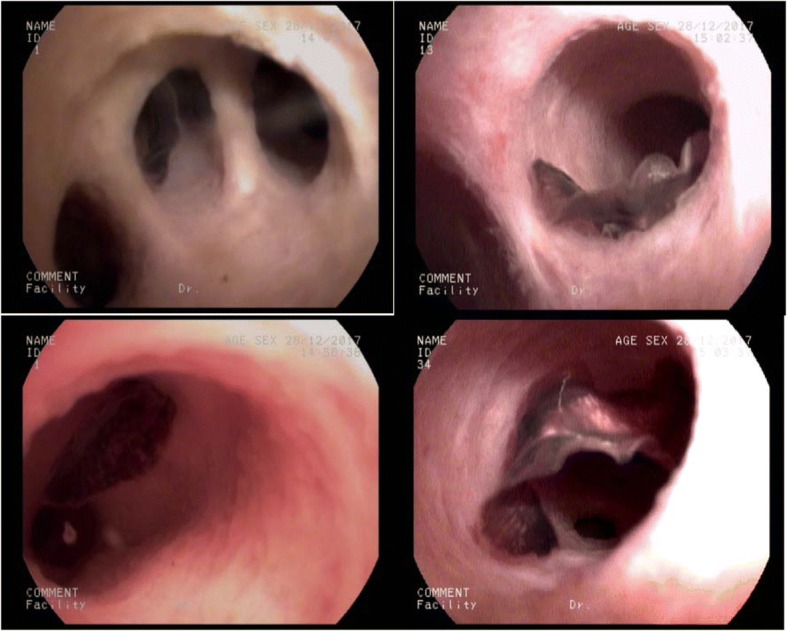
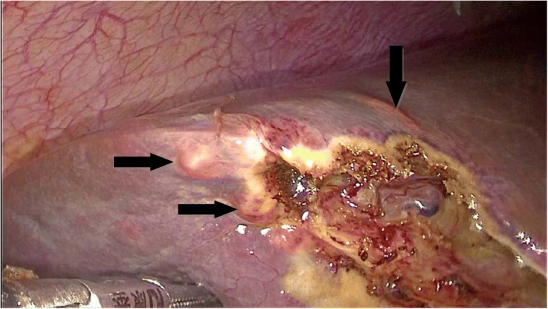
Case presentation: Two patients with habit of eating infected raw or undercooked freshwater fish were hospitalized due to right upper quadrant pain and jaundice. Magnetic resonance cholangiopancreatography (MRCP)/computed tomography (CT) detection indicated cholangiolithiasis and cholangiolithiasis with concurrent cholecystolithiasis, respectively. Fecal examinations were both negative for adult worms or eggs of parasites. However, adults of Clonrochis sinensis were detected within hepatobiliary tracts during laparoscopic cholecystectomy. Postoperative drainage and anthelmintic therapy contributed to complete recovery with good prognosis.
Conclusions: Clonorchiasis provokes cholangiolithiasis and cholecystolithiasis. Standardized treatments for these gallstone patients with concomitant clonorchiasis include surgical removal of the calculus, postoperative T tubule drainage and anthelmintic therapy. Serological test or polymerase chain reaction (PCR)-based approaches might be helpful for diagnosis of clonorchiasis when no eggs are found by stool microscopy. Public health promotion on ceasing to eat raw freshwater fish is essential for prevention and control of clonorchiasis.
Keywords: Clonorchiasis; Gallstone; Obstructive jaundice.
Conflict of interest statement
Ethics approval and consent to participate
All the clinical specimens were obtained with written consent and approved by the Fifth Affiliated Hospital of Sun Yat-sen University Ethics Committee, written consent was obtained from all patients involved in this study.
Consent for publication
The patients have signed the Consent form and agreed to publish this study and a copy of written Consent is available for the journal.
Competing interests
The authors declare that they have no competing interests.
Publisher’s Note
Springer Nature remains neutral with regard to jurisdictional claims in published maps and institutional affiliations.
Figures


Similar articles
-
Acute shock caused by Clonorchis sinensis infection: a case report.BMC Infect Dis. 2019 Nov 29;19(1):1014. doi: 10.1186/s12879-019-4644-5. BMC Infect Dis. 2019. PMID: 31783809 Free PMC article.
-
Obstructive jaundice caused by Clonorchiasis-associated duodenal papillitis: a case report.J Korean Med Sci. 2011 Jan;26(1):135-7. doi: 10.3346/jkms.2011.26.1.135. Epub 2010 Dec 22. J Korean Med Sci. 2011. PMID: 21218042 Free PMC article.
-
Clonorchis sinensis and clonorchiasis.Acta Trop. 2020 Mar;203:105309. doi: 10.1016/j.actatropica.2019.105309. Epub 2019 Dec 17. Acta Trop. 2020. PMID: 31862466 Review.
-
Painless Jaundice Caused by Clonorchis sinensis Infection: A Case Report.Korean J Parasitol. 2016 Jun;54(3):323-7. doi: 10.3347/kjp.2016.54.3.323. Epub 2016 Jun 30. Korean J Parasitol. 2016. PMID: 27417088 Free PMC article.
-
Clonorchiasis.Lancet. 2016 Feb 20;387(10020):800-10. doi: 10.1016/S0140-6736(15)60313-0. Epub 2015 Aug 21. Lancet. 2016. PMID: 26299184 Review.
Cited by
-
Unexpected discovery of Clonorchis sinensis by common bile duct exploration.Clin Case Rep. 2022 Feb 23;10(2):e05434. doi: 10.1002/ccr3.5434. eCollection 2022 Feb. Clin Case Rep. 2022. PMID: 35228877 Free PMC article.
-
Formation of non-cholesterol gallstones in populations within clonorchiasis endemic regions is closely related to Clonorchis sinensis infection.BMC Gastroenterol. 2025 May 8;25(1):345. doi: 10.1186/s12876-025-03939-3. BMC Gastroenterol. 2025. PMID: 40340656 Free PMC article.
-
Clonorchis sinensis and Echinostoma hortense detected by endoscopy and molecular characterization: two case reports and update on diagnosis.Front Med (Lausanne). 2025 Jan 22;11:1515539. doi: 10.3389/fmed.2024.1515539. eCollection 2024. Front Med (Lausanne). 2025. PMID: 39911668 Free PMC article.
-
Acute shock caused by Clonorchis sinensis infection: a case report.BMC Infect Dis. 2019 Nov 29;19(1):1014. doi: 10.1186/s12879-019-4644-5. BMC Infect Dis. 2019. PMID: 31783809 Free PMC article.
-
Clonorchiasis and opisthorchiasis: epidemiology, transmission, clinical features, morbidity, diagnosis, treatment, and control.Clin Microbiol Rev. 2024 Mar 14;37(1):e0000923. doi: 10.1128/cmr.00009-23. Epub 2024 Jan 3. Clin Microbiol Rev. 2024. PMID: 38169283 Free PMC article. Review.
References
Publication types
MeSH terms
Substances
Grants and funding
LinkOut - more resources
Full Text Sources
