

Danggui Buxue Tang, an ancient Chinese herbal decoction, protects β-amyloid-induced cell death in cultured cortical neurons
- PMID: 30621672
- PMCID: PMC6323849
- DOI: 10.1186/s12906-018-2411-6
Danggui Buxue Tang, an ancient Chinese herbal decoction, protects β-amyloid-induced cell death in cultured cortical neurons
Abstract
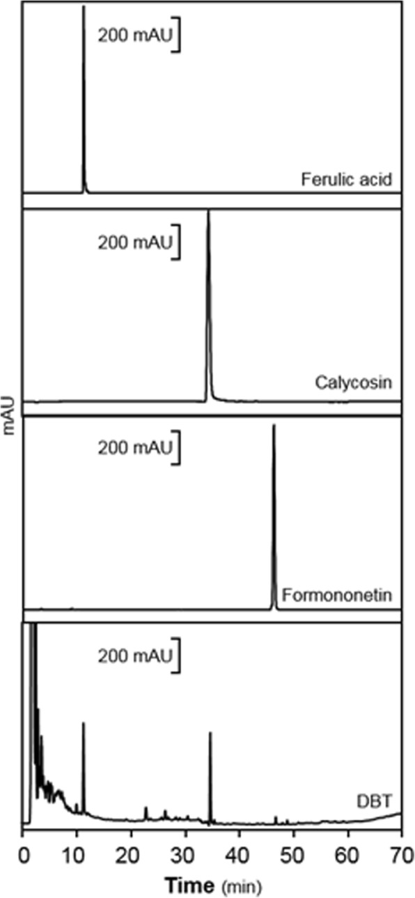
Background: Danggui Buxue Tang (DBT) is a historical Chinese herbal decoction, and which has more than 800 years of applications. This herbal decoction solely contains two materials: Astragali Radix (AR) and Angelicae Sinensis Radix (ASR) at a weight ratio of 5:1. Clinically, DBT aims to improve anemia syndrome. In complementary and alternative medicine theory, the cause of neurodegenerative disease is proposed to be related with anemia. In line to this notion, low levels of hemoglobin and red blood cell have been reported in patients suffering from Alzheimer's disease (AD), a chronic neurodegenerative disease caused by β-amyloid peptide (Aβ) accumulation. Therefore, we would like to probe the neuroprotective functions of this ancient herbal formula in vitro.
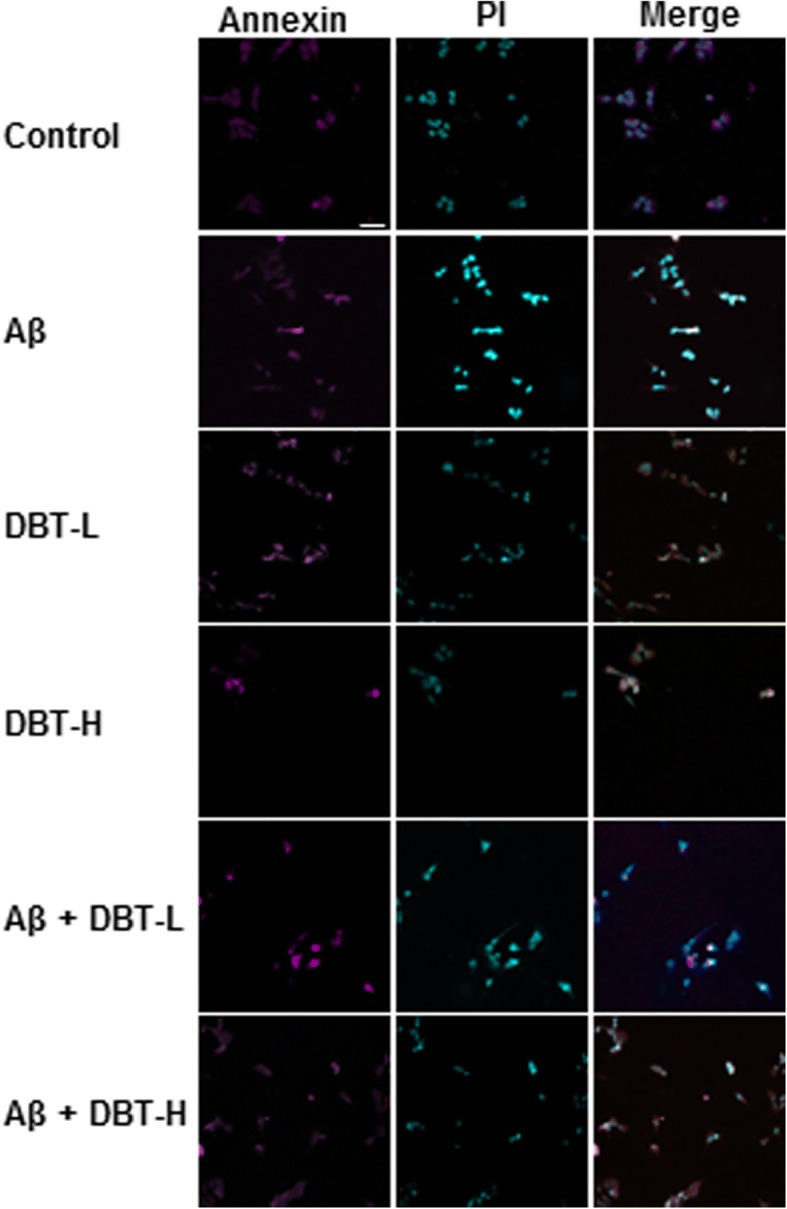
Method: The neuroprotective effects of DBT in the Aβ-induced cell death were detected in cultured cortical neurons by multiple techniques, i.e. confocal and western blot.
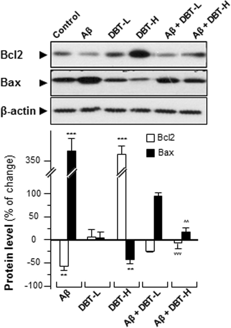
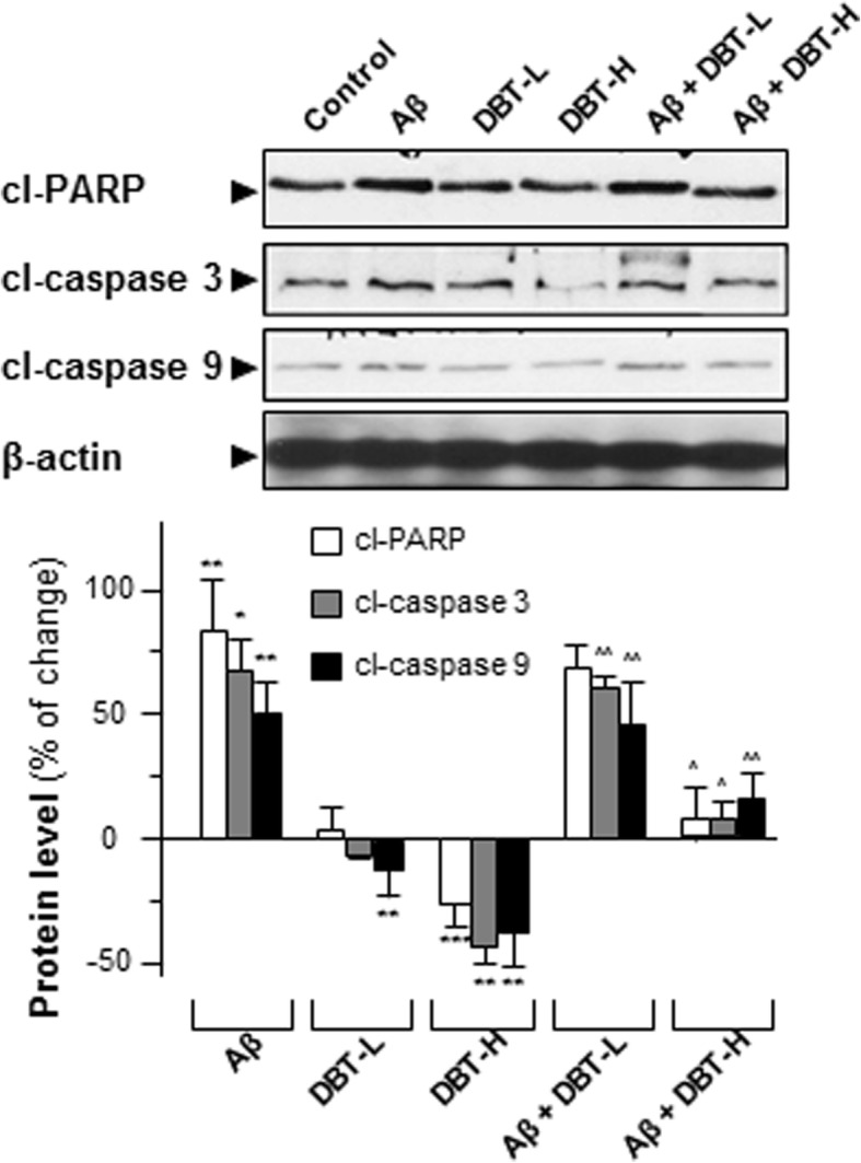
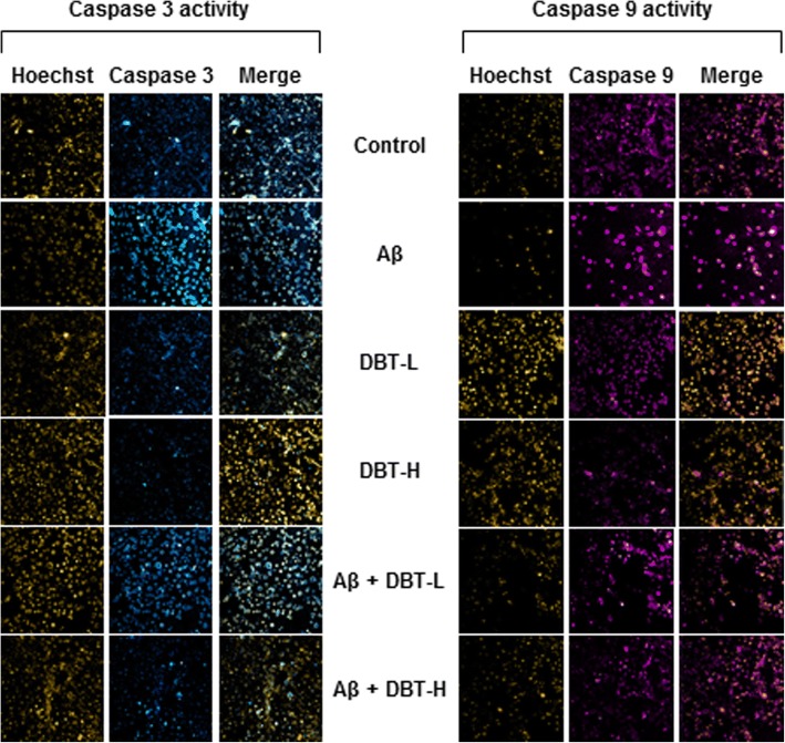
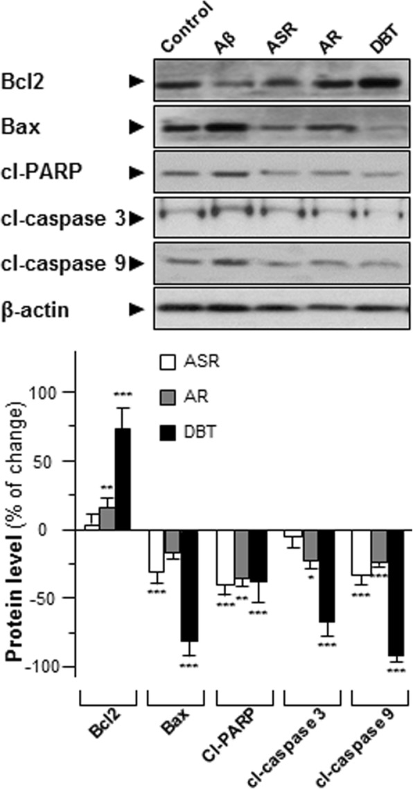
Results: In the cultures, application of DBT reduced Aβ-induced apoptosis rate in a dose-dependent manner. In Aβ-treated cortical neurons, the expression ratio of Bcl2 to Bax was altered by DBT. In parallel, application of DBT markedly suppressed the Aβ-induced expressions of apoptotic markers, i.e. cleaved-caspase 3/9 and PARP.
Conclusion: Taken these results, DBT shows promising protective effects against Aβ-induced stress or insult in cultured neurons.
Keywords: Alzhemier’s disease; Complementary and alternative medicine; Danggui Buxue Tang.
Conflict of interest statement
Ethics approval
The animal procedures were approved by The Animal Experimentation Ethics Committee of the Hong Kong University of Science and Technology (No. 17–283 for Animal Ethics Approval) and under the guidelines of “Principles of Laboratory Animal Care” (NIH publication No. DH/HA&P/8/2/3).
Consent for publication
Not applicable.
Competing interests
The authors declare that they have no competing interests.
Publisher’s Note
Springer Nature remains neutral with regard to jurisdictional claims in published maps and institutional affiliations.
Figures

Similar articles
-
Identification of Angelica oil as a suppressor for the biological properties of Danggui Buxue Tang: a Chinese herbal decoction composes of Astragali Radix and Angelica Sinensis Radix.J Ethnopharmacol. 2014 Jul 3;154(3):825-31. doi: 10.1016/j.jep.2014.05.007. Epub 2014 May 14. J Ethnopharmacol. 2014. PMID: 24837305
-
Can Hedysari Radix replace Astragali Radix in Danggui Buxue Tang, a Chinese herbal decoction for woman aliment?Phytomedicine. 2013 Sep 15;20(12):1076-81. doi: 10.1016/j.phymed.2013.04.016. Epub 2013 Jun 6. Phytomedicine. 2013. PMID: 23746954
-
Calycosin orchestrates the functions of Danggui Buxue Tang, a Chinese herbal decoction composing of Astragali Radix and Angelica Sinensis Radix: An evaluation by using calycosin-knock out herbal extract.J Ethnopharmacol. 2015 Jun 20;168:150-7. doi: 10.1016/j.jep.2015.03.033. Epub 2015 Mar 19. J Ethnopharmacol. 2015. PMID: 25796405 Review.
-
Danggui Buxue Tang (Astragali Radix and Angelicae Sinensis Radix) for menopausal symptoms: A review.J Ethnopharmacol. 2017 Mar 6;199:205-210. doi: 10.1016/j.jep.2017.01.044. Epub 2017 Feb 2. J Ethnopharmacol. 2017. PMID: 28163116 Review.
-
The expression of erythropoietin triggered by danggui buxue tang, a Chinese herbal decoction prepared from radix Astragali and radix Angelicae Sinensis, is mediated by the hypoxia-inducible factor in cultured HEK293T cells.J Ethnopharmacol. 2010 Oct 28;132(1):259-67. doi: 10.1016/j.jep.2010.08.029. Epub 2010 Aug 17. J Ethnopharmacol. 2010. PMID: 20723591
Cited by
-
Supplementation of dietary Angelica sinensis extracts to lactating Wuzhishan sows: effects on milk composition, immune function, milk-derived hormones, and related gene expression.Front Vet Sci. 2025 May 15;12:1524258. doi: 10.3389/fvets.2025.1524258. eCollection 2025. Front Vet Sci. 2025. PMID: 40444108 Free PMC article.
-
Exploration of the Danggui Buxue Decoction Mechanism Regulating the Balance of ESR and AR in the TP53-AKT Signaling Pathway in the Prevention and Treatment of POF.Evid Based Complement Alternat Med. 2021 Dec 30;2021:4862164. doi: 10.1155/2021/4862164. eCollection 2021. Evid Based Complement Alternat Med. 2021. PMID: 35003302 Free PMC article.
-
The Latest Research Advances of Danggui Buxue Tang as an Effective Prescription for Various Diseases: A Comprehensive Review.Curr Med Sci. 2022 Oct;42(5):913-924. doi: 10.1007/s11596-022-2642-0. Epub 2022 Oct 17. Curr Med Sci. 2022. PMID: 36245031 Review.
-
Dang-Gui-Bu-Xue decoction improves wound healing in diabetic rats by the activation of Notch signaling.Heliyon. 2024 Feb 20;10(5):e26711. doi: 10.1016/j.heliyon.2024.e26711. eCollection 2024 Mar 15. Heliyon. 2024. PMID: 38444491 Free PMC article.
-
Astragalus mongholicus Bunge (Fabaceae): Bioactive Compounds and Potential Therapeutic Mechanisms Against Alzheimer's Disease.Front Pharmacol. 2022 Jun 28;13:924429. doi: 10.3389/fphar.2022.924429. eCollection 2022. Front Pharmacol. 2022. PMID: 35837291 Free PMC article. Review.
References
-
- Flores KP, Blohowiak SE, Winzerling JJ, Georgieff MK, Kling PJ. The impact of erythropoietin and iron status on brain myelination in the newborn rat. J Neurosci Res. 2018. 10.1002/jnr.24243. - PubMed
MeSH terms
Substances
Grants and funding
LinkOut - more resources
Full Text Sources
Research Materials
