. 2019 Feb;21(2):206-215.
doi: 10.1016/j.neo.2018.12.005. Epub 2019 Jan 9.

MicroRNA-503-5p Inhibits the CD97-Mediated JAK2/STAT3 Pathway in Metastatic or Paclitaxel-Resistant Ovarian Cancer Cells

Affiliations

MicroRNA-503-5p Inhibits the CD97-Mediated JAK2/STAT3 Pathway in Metastatic or Paclitaxel-Resistant Ovarian Cancer Cells

Ga Bin Park et al. Neoplasia. 2019 Feb.

Abstract

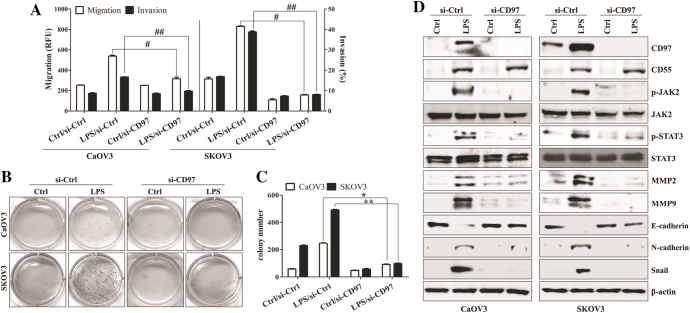
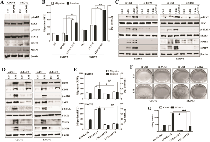
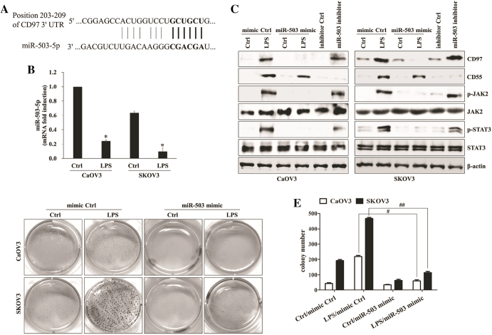
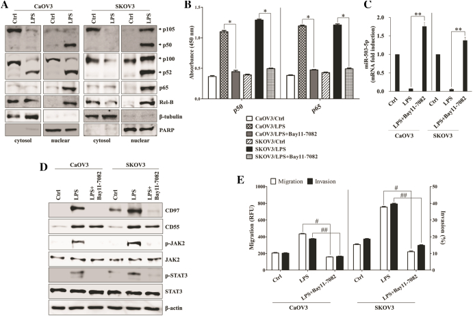
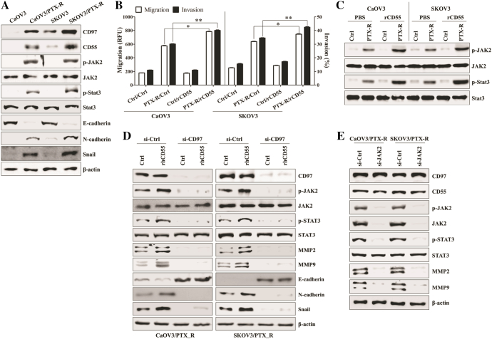
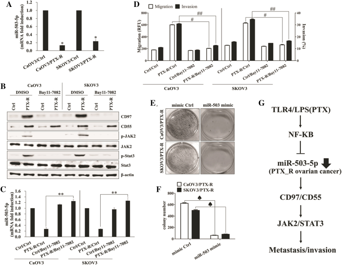
CD97 shows a strong relationship with metastasis and poor clinical outcome in various tumors, including ovarian cancer. The expression of CD97 in metastatic ovarian cancer cells was higher than that in primary ovarian cancer cells. Mature miRNAs are frequently de-regulated in cancer and incorporated into a specific mRNA, leading to post-transcriptional silencing. In this study, we investigated whether the miR-503-5p targeting of the CD97 3'-untranslated region (3'-UTR) contributes to ovarian cancer metastasis as well as the underlying mechanism regulating cancer progression. In LPS-stimulated or paclitaxel-resistant ovarian cancer cells, stimulation with recombinant human CD55 (rhCD55) of CD97 in ovarian cancer cells activated NF-κB-dependent miR-503-5p down-regulation and the JAK2/STAT3 pathway, consequently promoting the migratory and invasive capacity. Furthermore, restoration of miR-503-5p by transfection with mimics or NF-κB inhibitor efficiently blocked CD97 expression and the downstream JAK2/STAT3 signaling pathway. Target inhibition of JAK with siRNA also impaired colony formation and metastasis of LPS-stimulated and paclitaxel-resistant ovarian cancer cells. Taken together, these results suggest that high CD97 expression, which is controlled through the NF-κB/miR-503-5p signaling pathway, plays an important role in the invasive activity of metastatic and drug-resistant ovarian cancer cells by activating the JAK2/STAT3 pathway.


Figures

Figure 1
Figure 1
Knockdown of CD97 in LPS-stimulated ovarian cancer cells leads to decreased invasion and down-regulation of JAK2, STAT3, and MMP2/9. Cells (1.5 × 105/well) were seeded onto 6-well plates and grown overnight. Cells were transfected with siRNA against CD97 or control for 36 h and then treated with TLR4 agonist LPS (500 ng/ml) for 24 h. (A) The migratory activity and invasiveness of cells were detected by the tumor transendothelial migration assay kit and the BME cell invasion assay kit, respectively, as described in the Materials and Methods. #, P < .01. ##, P < .01. (B) Colony-forming assay. Cells were cultivated for 2 weeks in a 6-well plate with soft agar. After 2 weeks, the cells were stained with MTT solution. Colonies were counted after reaching at least 0.5 mm in diameter. (C) The graph shows the quantitative analysis of the colony-forming assay. *, P < .005. **, P < .005. Each value represents the mean ± SD of the three determinations. (D) Total cell lysates were immunoblotted with the indicated antibodies. β-actin was used as a loading control. The results are representative of three independent experiments.
Figure 2
Figure 2
CD97/CD55 interaction induces JAK2/STAT3-mediated metastasis of LPS-stimulated ovarian cancer cells. (A) Cells (1.5 × 105/well) were cultured with recombinant human CD55 (1 μg/ml) for 24 h. Total cell lysates were immunoblotted with the indicated antibodies. (B) Cells (1.5 × 105/well) were cultured with recombinant human CD55 (1 μg/ml) and LPS (500 ng/ml) for 24 h. The migratory activity and invasiveness of cells were detected by the tumor transendothelial migration assay kit and the BME cell invasion assay kit, respectively, as described in the Materials and Methods. *, P < .01. **, P < .01. (C) Cells (1.5 × 105/well) were seeded onto 6-well plates and grown overnight. Cells were transfected with siRNA against CD97 or control for 36 h and then treated with recombinant human CD55 (1 μg/ml) and LPS (500 ng/ml) for 24 h. Total cell lysates were immunoblotted with the indicated antibodies. (D-G) Cells (1.5 × 105/well) were seeded onto 6-well plates and grown overnight. Cells were transfected with siRNA against JAK2 or control for 36 h and then treated with LPS (500 ng/ml) for 24 h. (D) Total cell lysates were immunoblotted with the indicated antibodies. β-actin was used as a loading control. (E) The migratory activity and invasiveness of cells were detected by the tumor transendothelial migration assay kit and the BME cell invasion assay kit, respectively, as described in the Materials and Methods. #, P < .01. ##, P < .01. (F) Colony-forming assay. Cells were cultivated for 2 weeks in a 6-well plate with soft agar. After 2 weeks, the cells were stained with MTT solution. Colonies were counted after reaching at least 0.5 mm in diameter. (G) The graph shows the quantitative analysis of the colony-forming assay. ♠, P < .005. ♠♠, P < .005. Each value represents the mean ± SD of the three determinations. The results are representative of three independent experiments.
Figure 3
Figure 3
miR-503-5p binds directly to the 3’UTRs of CD97 and LPS-induced miR-503-5p down-regulation enhances invasion. (A) A schematic of the putative miR-503-5p binding sites on CD97 3′-UTR. (B) Quantitative real-time RT-PCR (qPCR) was performed to determine the relative expression of miR-503-5p in LPS-treated ovarian cancer cells. *, P < .05. (C-E) Cells (1.5 × 105/well) were seeded onto 6-well plates and grown overnight. Cells were transfected with miR-503-5p mimic or mimic control for 36 h and then treated with LPS (500 ng/ml) for 24 h. Some cells were transfected with miR-503-5p inhibitor or inhibitor control for 48 h. (C) Total cell lysates were immunoblotted with the indicated antibodies. β-actin was used as a loading control. (D) Colony-forming assay. Cells were cultivated for 2 weeks in a 6-well plate with soft agar. After 2 weeks, the cells were stained with MTT solution. Colonies were counted after reaching at least 0.5 mm in diameter. (E) The graph shows the quantitative analysis of the colony-forming assay. #, P < .01. ##, P < .01. Each value represents the mean ± SD of the three determinations. The results are representative of three independent experiments.
Figure 4
Figure 4
NF-κB-mediated miR-503-5p down-regulation increases the CD97-induced JAK2/STAT3 expression and invasion. (A) Cells (1.5 × 105/well) were cultured with LPS (500 ng/ml) for 24 h. Cytosolic extracts or nuclear extracts were analyzed by Western blotting using the indicated antibodies. A nuclear marker, PARP, and cytosol marker, β-tubulin, were used to verify the purity of each fraction. Fractionation was performed as described in the Materials and Methods. (B-E) Cells (1.5 × 105/well) were pretreated with Bay 11–7082 (5 μM) for 1 h and then treated with LPS (500 ng/ml) for 24 h. (B) ELISA measured NF-κB DNA-binding activity in nuclear extracts. Transcription factor NF-κB p50 combo and p65 combo (in Kit) served as positive controls for NF-κB activity. ELISA results are expressed as relative absorbance. *, P < .01. Data represent the mean ± SD of the three independent experiments. (C) QPCR was performed to determine the relative expression of miR-503-5p in LPS and Bay 11–7082 (5 μM)-treated ovarian cancer cells. **, P < .05. (D) Total cell lysates were immunoblotted with the indicated antibodies. β-actin was used as a loading control. (E) The migratory activity and invasiveness of cells were detected by the tumor transendothelial migration assay kit and the BME cell invasion assay kit, respectively, as described in the Materials and Methods. #, P < .005. ##, P < .005. Each value represents the mean ± SD of the three determinations. The results are representative of three independent experiments.
Figure 5
Figure 5
CD97/CD55 interaction elicits JAK2/STAT3-mediated metastasis of paclitaxel-resistant ovarian cancer cells. (A) Total lysates of PTX-sensitive or PTX-resistant cells were collected and immunoblotted with the indicated antibodies. (B, C) Cells (1.5 × 105/well) were cultured with recombinant human CD55 (1 μg/ml) for 24 h. (B) The migratory activity and invasiveness of cells were detected by the tumor transendothelial migration assay kit and the BME cell invasion assay kit, respectively, as described in the Materials and Methods. *, P < .005. **, P < .005. Each value represents the mean ± SD of the three determinations. (C) Total cell lysates were collected and immunoblotted with the indicated antibodies. (D) Cells (1.5 × 105/well) were seeded onto 6-well plates and grown overnight. Cells were transfected with siRNA against JAK2 or control for 48 h. Total cell lysates were collected and immunoblotted with the indicated antibodies. β-actin was used as a loading control. The results are representative of three independent experiments.
Figure 6
Figure 6
The level of miR-503-5p modulates CD97-mediated metastasis of paclitaxel-resistant ovarian cancer cells. (A) qPCR was performed to determine the relative expression of miR-503-5p in PTX-sensitive or PTX-resistant ovarian cancer cells. *, P < .05. (B-D) Cells (1.5 × 105/well) were treated with Bay 11–7082 (5 μM) for 1 h, washed out, and then cultured for 24 h. (B) Total cell lysates were immunoblotted with the indicated antibodies. β-actin was used as a loading control. (C) the levels of miR-503-5p in PTX-sensitive or PTX-resistant ovarian cancer cells were measured using qPCR. **, P < .05. (D) The migratory activity and invasiveness of cells were detected by the tumor transendothelial migration assay kit and the BME cell invasion assay kit, respectively, as described in the Materials and Methods. #, P < .005. ##, P < .005. (E, F) Cells (1.5 × 105/well) were seeded onto 6-well plates and grown overnight. Cells were transfected with miR-503-5p mimic or mimic control for 48 h. (E) Colony-forming assay. Cells were cultivated for 2 weeks in a 6-well plate with soft agar. After 2 weeks, cells were stained with MTT solution. Colonies were counted after reaching at least 0.5 mm in diameter. (F) The graph shows the quantitative analysis of the colony-forming assay. ♠, P < .01. Each value represents the mean ± SD of the three determinations. The results are representative of three independent experiments. (G) Schematic diagram of the intracellular signaling pathway in TLR4 agonist-treated human ovarian cancer cells. TLR4 stimulation triggers NF-κB activation in ovarian cancer cells. Subsequent suppression of miR-503-5p results in the increase of CD97 and CD55 expression. The interaction of CD97 and CD55 leads to JAK2/STAT3-mediated invasion in LPS-treated ovarian cancer cells. Our findings suggest that ovarian cancer cells exposure to LPS plays an important role in cancer metastasis through the modulation of miR-503-5p and the regulation of CD97/CD55.

Similar articles

Cited by

References

    1. Eichler W, Hamann J, Aust G. Expression characteristics of the human CD97 antigen. Tissue Antigens. 1997;50:429–438. - PubMed
    1. Yona S, Lin HH, Siu WO, Gordon S, Stacey M. Adhesion-GPCRs: emerging roles for novel receptors. Trends Biochem Sci. 2008;33:491–500. - PubMed
    1. Aust G, Steinert M, Schütz A, Boltze C, Wahlbuhl M, Hamann J, Wobus M. CD97, but not its closely related EGF-TM7 family member EMR2, is expressed on gastric, pancreatic, and esophageal carcinomas. Am J Clin Pathol. 2002;118:699–707. - PubMed
    1. Safaee M, Clark AJ, Ivan ME, Oh MC, Bloch O, Sun MZ, Oh T, Parsa AT. CD97 is a multifunctional leukocyte receptor with distinct roles in human cancers. Int J Oncol. 2013;43:1343–1350. - PubMed
    1. Han SL, Xu C, Wu XL, Li JL, Liu Z, Zeng QQ. The impact of expressions of CD97 and its ligand CD55 at the invasion front on prognosis of rectal adenocarcinoma. Int J Color Dis. 2010;25:695–702. - PubMed

Publication types

MeSH terms