. 2019 Jan 8;10(1):e02506-18.
doi: 10.1128/mBio.02506-18.

RNA Structure Duplication in the Dengue Virus 3' UTR: Redundancy or Host Specificity?

Affiliations

RNA Structure Duplication in the Dengue Virus 3' UTR: Redundancy or Host Specificity?

Luana de Borba et al. mBio. .

Abstract

Flaviviruses include a diverse group of medically important viruses that cycle between mosquitoes and humans. During this natural process of switching hosts, each species imposes different selective forces on the viral population. Using dengue virus (DENV) as model, we found that paralogous RNA structures originating from duplications in the viral 3' untranslated region (UTR) are under different selective pressures in the two hosts. These RNA structures, known as dumbbells (DB1 and DB2), were originally proposed to be enhancers of viral replication. Analysis of viruses obtained from infected mosquitoes showed selection of mutations that mapped in DB2. Recombinant viruses carrying the identified variations confirmed that these mutations greatly increase viral replication in mosquito cells, with low or no impact in human cells. Use of viruses lacking each of the DB structures revealed opposite viral phenotypes. While deletion of DB1 reduced viral replication about 10-fold, viruses lacking DB2 displayed a great increase of fitness in mosquitoes, confirming a functional diversification of these similar RNA elements. Mechanistic analysis indicated that DB1 and DB2 differentially modulate viral genome cyclization and RNA replication. We found that a pseudoknot formed within DB2 competes with long-range RNA-RNA interactions that are necessary for minus-strand RNA synthesis. Our results support a model in which a functional diversification of duplicated RNA elements in the viral 3' UTR is driven by host-specific requirements. This study provides new ideas for understanding molecular aspects of the evolution of RNA viruses that naturally jump between different species.IMPORTANCE Flaviviruses constitute the most relevant group of arthropod-transmitted viruses, including important human pathogens such as the dengue, Zika, yellow fever, and West Nile viruses. The natural alternation of these viruses between vertebrate and invertebrate hosts shapes the viral genome population, which leads to selection of different viral variants with potential implications for epidemiological fitness and pathogenesis. However, the selective forces and mechanisms acting on the viral RNA during host adaptation are still largely unknown. Here, we found that two almost identical tandem RNA structures present at the viral 3' untranslated region are under different selective pressures in the two hosts. Mechanistic studies indicated that the two RNA elements, known as dumbbells, contain sequences that overlap essential RNA cyclization elements involved in viral RNA synthesis. The data support a model in which the duplicated RNA structures differentially evolved to accommodate distinct functions for viral replication in the two hosts.

Keywords: RNA virus; RNA virus evolution; RNA-RNA interactions; dengue virus; flavivirus; host adaptation; viral RNA structures.


Figures

FIG 1
FIG 1
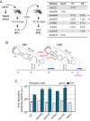
DENV adaptive mutations associated with replication in mosquitoes. (A) (Left) Experimental design of DENV adaptation in adult mosquitoes. (Right) Mutations identified in the input and passages P1 and P2. Red stars indicate mutations that showed increased frequencies from P1 to P2. (B) Location of adaptive mutations in the viral 3′ UTR. The secondary structures of the two DBs are shown. Nucleotide variations are indicated in red. (C) Replication of recombinant reporter DENVs carrying the identified mutations. The plot shows viral replication measured by luciferase activity as a function of time post-RNA transfection into mosquito cells. The luciferase values (in arbitrary units [a.u.]) are means ± standard deviations (representative results of three independent experiments). *, P < 0.05; ***, P < 0.001.
FIG 2
FIG 2
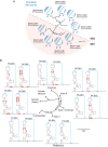
Sequence and structure analysis of the two DB elements. (A) Fan dendrogram indicating the distance of DB structures from different DENV serotypes. The corresponding circle plot for each sequence is shown with arcs denoting base pairs. (B) Comparison of the natural sequence variability of DB1 and DB2 for different DENV2 genotypes. Asian I genotype was used as a reference. Variability of the DB sequences is indicated in red. The size bar indicates number of nucleotide substitution per site.
FIG 3
FIG 3
Divergent functions of the two DB structures. (A) Schematic representation of the DENV genome showing the RNA structures in the 5′ and 3′ UTRs. (B and E) Replication of DENV reporter mutants carrying deletions of individual DBs (ΔDB1 and ΔDB1) or both (ΔDB1-2) in mosquito and human cells, respectively. Plots show Renilla luciferase activity as a function of time post-RNA transfection. (C and D) Replication of WT DENV or mutants with DB deletions, using the original infectious DENV2 16681 clone. Luciferase values are means ± standard deviations (representative results from three independent experiments). (C) RNA accumulation. The plot indicates number of viral genome copies as a function of time in mosquito cells. (D) Production of infectious particles is expressed as PFU/ml. (F) Impact of the deletion of each DB on viral replication in mosquito and human cells. (G) Accumulation of sfRNAs in mosquito cells. Shown is a Northern blot using a radiolabeled probe that recognizes the viral RNA 3′ UTR. Different sfRNA species are indicated on the left (sfRNA1-4 and sfRNA1′). (H) Schematic representation of the del30 deleted sequence in DB2 and infectious particles produced in mosquito cells (top) and replication of the DENV del30 reporter mutant in mosquito and human cells along with the WT. Luciferase values are means ± standard deviations (representative results from three independent experiments). (I) Accumulation of gRNA (genomic RNA) and sfRNAs in mosquito cells. Shown is a Northern blot using a radiolabeled probe that recognizes the viral RNA 3′ UTR for sfRNA and NS2B for gRNA. Different sfRNA species separated by PAGE are indicated on the left. Asterisks indicate different sizes of sfRNA1 because of the DB deletions. The plot on the right shows the ratio of sfRNA to gRNA for each mutant.
FIG 4
FIG 4
Mutually exclusive local and long-range RNA interactions. Shown is a schematic representation of linear and circular conformations of the DENV genome. Cyclization elements are indicated: 5′ and 3′ UAR (blue), 5′ and 3′ CS (purple), 5′ and 3′ DAR (gray), and C1-DB1 (red) in both structures (top linear and bottom circular). DB2 is indicated in green. In boxes, the sequences of pseudoknot (PK-IV) that overlap the 3′ CS are shown for both forms—linear (PK-IV) and circular (5′-3′ CS).
FIG 5
FIG 5
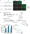
DB2 sequence negatively regulates genome cyclization. (A) Immunofluorescence of mosquito cells transfected with RNA from WT and recombinant viruses carrying the mutations Mut Cyc+ and Mut Cyc in the 5′-3′ UAR sequence, as indicated. (B) Schematic representation of mutations identified in the recovered viruses. In the case of Mut Cyc+, the location of the mutations in linear and circular forms of the RNA is shown. In the case of Mut Cyc, the location of mutations in the circular form is shown, indicating both the complementary region and disruption of DB2 PK. (C) (Left) Design of mutation to disrupt the DB2 PK (MutPK). (Right) Replication of DENV reporter mutants in mosquito cells. Luciferase activity measurements as a function of time are shown for each case. The luciferase values are means ± standard deviations (representative results from three independent experiments). **, P < 0.01.
FIG 6
FIG 6
Mapping single nucleotide variations in the viral 3′ UTR from intrahost analysis in DENV1-infected mosquitoes (A) Distribution along the viral 3′ UTR of mutations identified in an intrahost analysis using A. aegypti mosquitoes. The plot represents the frequency of mutations identified indicated in the y axis and the position in a reference DENV1 genome (GenBank accession no. HG316481) shown in the x axis. Colors indicate each of the defined RNA structures along the 3′ UTR, as indicated on the right. Sequence variations detected in viruses included in the blood meal are also shown in the plot by open circles and on the right, indicating the position, nucleotide change, and frequency for each case. For SNVs detected in several samples, only the highest frequency is indicated. (B) Location of the mutations identified with frequencies above 10% on the secondary structure of the DENV1 3′ UTR. As a reference DENV1, GenBank accession no. HG316481 was used. Arrows indicate the position of the substitutions, including the frequency, the nucleotide change, and the source—midgut (MG) or salivary gland (SG). The green circle indicates a mutation found at a frequency of 99% that disrupts DB2-PK formation.
FIG 7
FIG 7
Conserved distribution of DB PK and cyclization elements in the genomes of mosquito-borne flaviviruses (MBFVs). Dumbbell structures of selected viruses from each subgroup of MBFV are shown. Locations and complementary bases that form PKs are indicated in blue. YFV and ZIKV form ΨDBs with PKs that are indicated in gray. Complementary sequences between the 5′ and 3′ ends of the genome that involve DB PKs are shown (5′-3′ interactions). Estimated P values show the reliability of indicated long-range RNA-RNA interactions, which were compared with a randomly sampled alignment. The distance tree was drawn using the neighbor-joining method of all complete genome sequences for each virus available in GenBank.

References

    1. Bhatt S, Gething PW, Brady OJ, Messina JP, Farlow AW, Moyes CL, Drake JM, Brownstein JS, Hoen AG, Sankoh O, Myers MF, George DB, Jaenisch T, Wint GR, Simmons CP, Scott TW, Farrar JJ, Hay SI. 2013. The global distribution and burden of dengue. Nature 496:504–507. doi:10.1038/nature12060. - DOI - PMC - PubMed
    1. Mir D, Delatorre E, Bonaldo M, Lourenco-de-Oliveira R, Vicente AC, Bello G. 2017. Phylodynamics of yellow fever virus in the Americas: new insights into the origin of the 2017 Brazilian outbreak. Sci Rep 7:7385. doi:10.1038/s41598-017-07873-7. - DOI - PMC - PubMed
    1. Iglesias N, Byk L, Gamarnik A. 2014. Molecular biology of dengue virus, p 334–364. In Gubler DJ, Ooi EE, Vasudevan S, Farrar J (ed), Dengue and dengue hemorrhagic fever, 2nd ed CABI, Wallingford, United Kingdom.
    1. Manokaran G, Finol E, Wang C, Gunaratne J, Bahl J, Ong EZ, Tan HC, Sessions OM, Ward AM, Gubler DJ, Harris E, Garcia-Blanco MA, Ooi EE. 2015. Dengue subgenomic RNA binds TRIM25 to inhibit interferon expression for epidemiological fitness. Science 350:217–221. doi:10.1126/science.aab3369. - DOI - PMC - PubMed
    1. Villordo SM, Filomatori CV, Sanchez-Vargas I, Blair CD, Gamarnik AV. 2015. Dengue virus RNA structure specialization facilitates host adaptation. PLoS Pathog 11:e1004604. doi:10.1371/journal.ppat.1004604. - DOI - PMC - PubMed

Publication types

LinkOut - more resources