. 2019 Jan 8;10(1):88.
doi: 10.1038/s41467-018-08007-x.

Inward- and outward-facing X-ray crystal structures of homodimeric P-glycoprotein CmABCB1

Affiliations

Inward- and outward-facing X-ray crystal structures of homodimeric P-glycoprotein CmABCB1

Atsushi Kodan et al. Nat Commun. .

Abstract

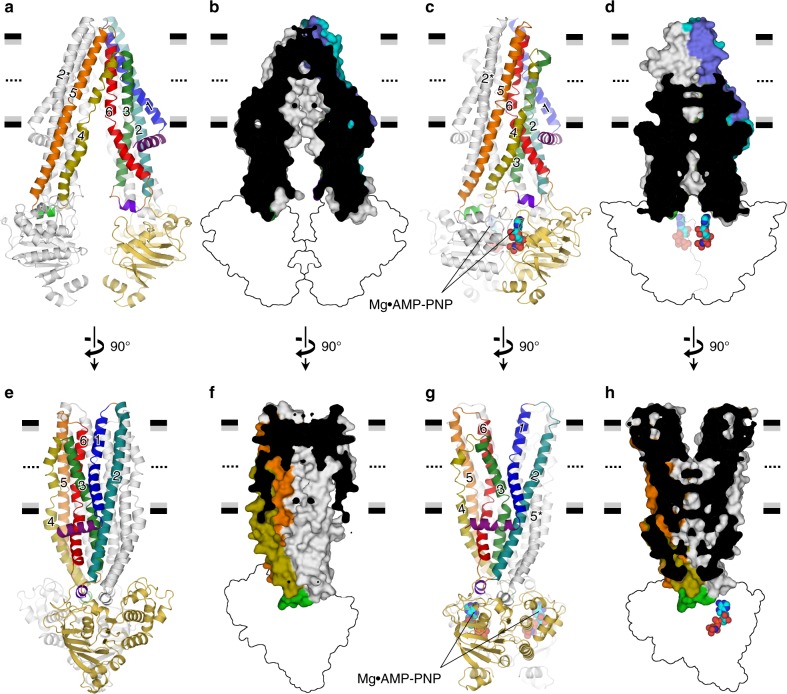
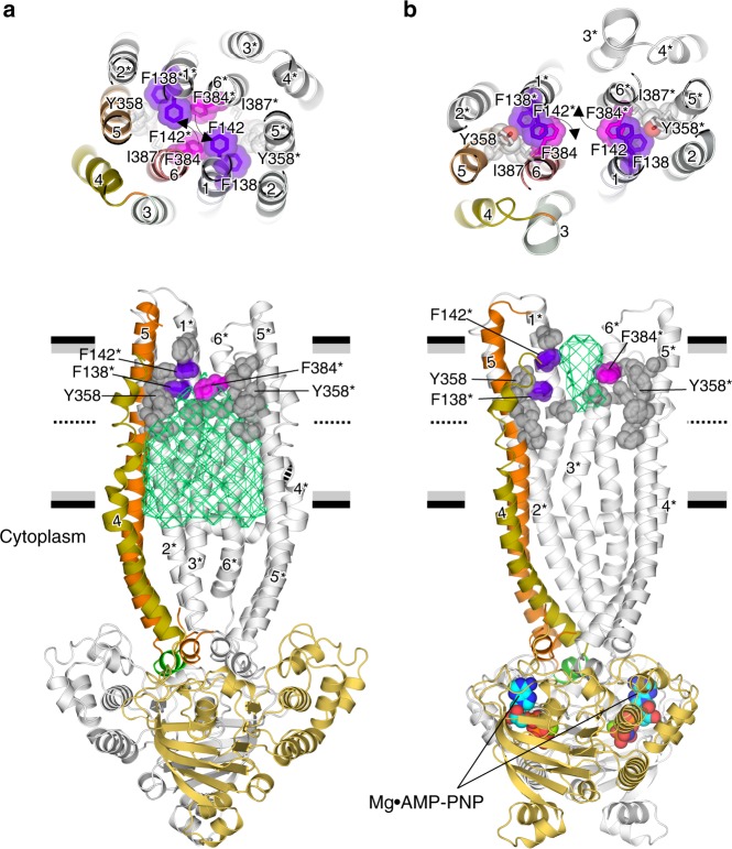
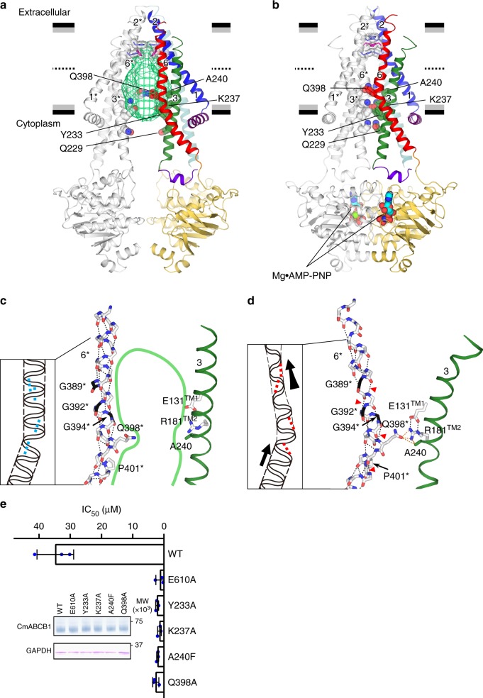
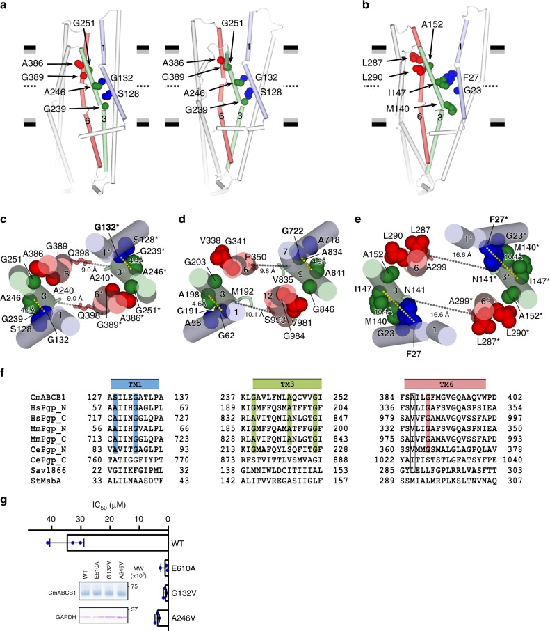
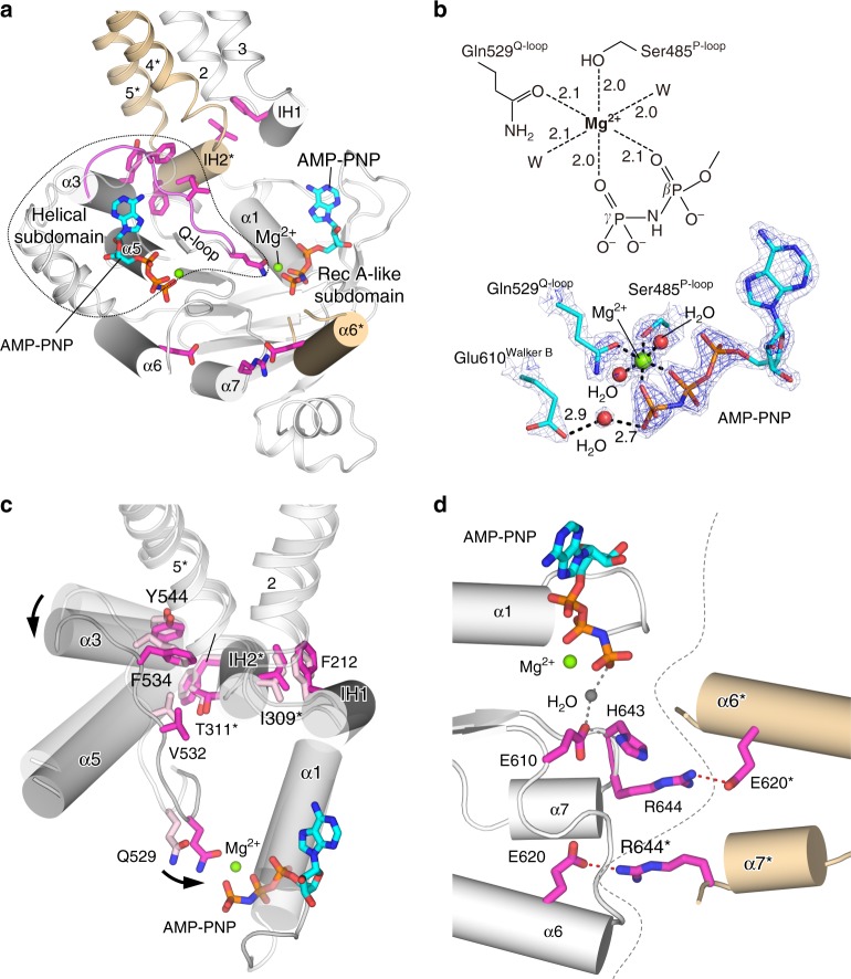
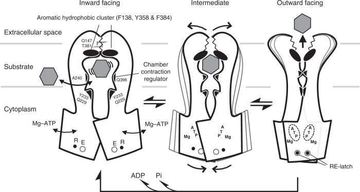
P-glycoprotein extrudes a large variety of xenobiotics from the cell, thereby protecting tissues from their toxic effects. The machinery underlying unidirectional multidrug pumping remains unknown, largely due to the lack of high-resolution structural information regarding the alternate conformational states of the molecule. Here we report a pair of structures of homodimeric P-glycoprotein: an outward-facing conformational state with bound nucleotide and an inward-facing apo state, at resolutions of 1.9 Å and 3.0 Å, respectively. Features that can be clearly visualized at this high resolution include ATP binding with octahedral coordination of Mg2+; an inner chamber that significantly changes in volume with the aid of tight connections among transmembrane helices (TM) 1, 3, and 6; a glutamate-arginine interaction that stabilizes the outward-facing conformation; and extensive interactions between TM1 and TM3, a property that distinguishes multidrug transporters from floppases. These structural elements are proposed to participate in the mechanism of the transporter.


Conflict of interest statement

The authors declare no competing interests.

Figures

Fig. 1
Fig. 1
Apo inward-facing and Mg2+•AMP-PNP-bound outward-facing structures of QTA CmABCB1. Anterior (ad) and lateral (e, f) views of the QTA CmABCB1 structures in the inward- (a, b, e, f) and outward-facing (c, d, g, h) conformations are shown as cartoon representations (a, c, e, g) or as a cutaway surface representations of the TMDs with the interior shown in black (b, d, f, h). One subunit is colored, and the other is shown in gray. Secondary structure elements and amino acid residues belonging to the other subunit are indicated by asterisks in all figures. Horizontal black and gray bars represent the expected positions of the hydrophilic and hydrophobic surfaces of the lipid membrane, respectively. In b, f, d, h, the cross-section passes through a crystallographic twofold axis. NBDs are shown as outlines for simplicity. In c, d, g, h, bound Mg2+•AMP-PNP molecules at NBDs are shown as spheres
Fig. 2
Fig. 2
Structural changes forming a substrate pathway from the inner side to the extracellular side. Rearrangement of side chains, primarily in TM1, TM6, TM1*, and TM6*, between inward- (a) and outward-facing (b) conformations of QTA CmABCB1. (Upper panels) Top views of the extracellular gate, showing transition of residues lining TM1, TM6, TM1*, and TM6*. (Lower panels) Lateral views of both structures. For clarity, several TM helices (TM1, TM2, TM3, and TM6) are omitted. One subunit is colored in orange, and the other is shown in gray. The inner chamber in the inward-facing conformation is shown as a green mesh (a). In b, bound Mg2+•AMP-PNP molecules at NBDs are shown as spheres. Horizontal black and gray bars represent the expected positions of the hydrophilic and hydrophobic surfaces of the lipid membrane, respectively
Fig. 3
Fig. 3
Large change in the chamber volume. a, b The inside anterior views of inward- (a) and outward-facing (b) conformations of QTA CmABCB1. For clarity, TM4, TM5, TM4*, and TM5* shown in Fig. 1a, c are omitted here. Residues forming close inter-subunit contacts on the cytosolic side in the outward-facing conformation are shown as spheres. The inner chamber in the inward-facing conformation is shown as a green mesh (a). One subunit is shown in color, and the other is shown in gray. Horizontal black and gray bars represent the expected positions of the hydrophilic and hydrophobic surfaces of the lipid membrane, respectively. In b, bound Mg2+•AMP-PNP molecules at NBDs are shown as spheres. c, d Close-up of TM6* and TM3 in inward- (a) and outward-facing (b) conformations viewed parallel to the membrane. Intra-helical interactions within the main chains of TM6 are shown as dashed lines. In c, the internal large cavity facing the intracellular side in the inward-facing conformation is outlined in green. In d, TM3 and the side chains of residues interacting with Gln398 are shown as ribbon and sticks, respectively. Polar and van der Waals interactions are shown as black and orange dashed lines, respectively. Non-α-helical hydrogen bonds in TM6 are indicated by red arrowheads. The stretching directions of TM6 from the inward- to outward-facing conformations are shown as thick black arrows in the schematic. e Bar graph, overlaid with the actual data points, shows IC50 for growth inhibition in a rhodamine 6G susceptibility assay using S. cerevisiae AD1-8u cells. Error bars indicate standard deviation (n = 3). Cells expressing the ATPase-deficient mutant E610A served as controls. Inset shows the amounts of mutant and WT CmABCB1 expressed in AD1-8u cells, as determined by western blotting. Uncropped images of the blots are shown in Supplementary Fig. 6
Fig. 4
Fig. 4
TM joints characterizing the outward-facing structure of P-gp. a, b Inter-helical interactions of TM1−TM3 and TM3−TM6 of inward- (a, left) and outward-facing (a, right) conformations of QTA CmABCB1, and comparison with outward-facing structure of Sav1866 (b). Residues serving as TM joints in CmABCB1 and corresponding residues in Sav1866 are shown as spheres. Only the TMD of one subunit is shown for simplicity. ce Arrangement of TM joints of outward-facing QTA CmABCB1 (c) and human P-gp (d) viewed from the extracellular side and comparison with Sav1866 (e). TM1 (TM1* or TM7), TM3 (TM3* or TM9), and TM6 (TM6* or TM12) are shown as cylinders. Residues serving as TM joints in CmABCB1 and the corresponding residues in human P-gp and Sav1866 are shown as spheres. f Local sequence alignment of the TM joints consisting of TM1, TM3, and TM6. Conserved Gly and other residues with small side chains, such as Ala, and Ser, are highlighted. g Bar graph, overlaid with the actual data points, shows IC50 for growth inhibition in rhodamine 6G susceptibility assay using S. cerevisiae AD1-8u cells. Error bars indicate standard deviation (n = 3). Cells expressing the ATPase-deficient mutant E610A served as controls. Inset shows the amounts of mutant and WT CmABCB1 expressed in AD1-8u cells, as determined and analyzed by western blotting. Uncropped images of the blots are shown in Supplementary Fig. 6
Fig. 5
Fig. 5
Coupling between NBD−TMD and NBD−NBD* in the outward-facing conformation of QTA CmABCB1. a NBD bound to Mg2+•AMP-PNP in a subunit of QTA CmABCB1, viewed parallel to the membrane. The NBD and TM2−IH1−TM3 of one subunit are shown in white, except for Q-loop-α3, shown in magenta; TM4*−IH2*−TM5* and α6* of the other subunit are shown in orange. The key residues for coupling between NBD−TMD and NBD−NBD* are shown as magenta sticks. b Octahedral coordination of Mg2+. Light blue and blue meshes represent the 2Fo−Fc map contoured at 2.7 and 4.5 sigma, respectively. Polar interactions are shown as dashed lines. c Superposition of the NBDs in the outward- (gray) and inward-facing (white) conformations, based on the RecA-like subdomain. The direction of movement of α3 and Gln529 between the inward- and outward-facing conformations are shown as arrows. Residues contributing to the coupling of movement between the NBD and TMD are shown as sticks. d Interaction between NBDs stabilized by RE-latch. The salt bridges in RE-latch are shown as red dashed lines, and the boundary between the two NBDs is shown as gray dashed lines
Fig. 6
Fig. 6
Proposed model for transport mechanism of CmABCB1. Schematic drawing of conformational change between the inward- (left) and the outward-facing (right) CmABCB1. An expected intermediate state is depicted in the center panel. The characteristic devices indispensable for the mechanics of transport are represented

References

    1. Ueda K, Cardarelli C, Gottesman MM, Pastan I. Expression of a full-length cDNA for the human “MDR1” gene confers resistance to colchicine, doxorubicin, and vinblastine. Proc. Natl. Acad. Sci. USA. 1987;84:3004–3008. doi: 10.1073/pnas.84.9.3004. - DOI - PMC - PubMed
    1. Schinkel AH, Jonker JW. Mammalian drug efflux transporters of the ATP binding cassette (ABC) family: an overview. Adv. Drug Deliv. Rev. 2003;55:3–29. doi: 10.1016/S0169-409X(02)00169-2. - DOI - PubMed
    1. Szakacs G, Paterson JK, Ludwig JA, Booth-Genthe C, Gottesman MM. Targeting multidrug resistance in cancer. Nat. Rev. Drug Discov. 2006;5:219–234. doi: 10.1038/nrd1984. - DOI - PubMed
    1. Seelig A. A general pattern for substrate recognition by P-glycoprotein. Eur. J. Biochem. 1998;251:252–261. doi: 10.1046/j.1432-1327.1998.2510252.x. - DOI - PubMed
    1. Szakacs G, Varadi A, Ozvegy-Laczka C, Sarkadi B. The role of ABC transporters in drug absorption, distribution, metabolism, excretion and toxicity (ADME-Tox) Drug Discov. Today. 2008;13:379–393. doi: 10.1016/j.drudis.2007.12.010. - DOI - PubMed

Publication types

MeSH terms