. 2018 Dec 21:12:495.
doi: 10.3389/fncel.2018.00495. eCollection 2018.

Fractalkine/CX3CR1 Contributes to Endometriosis-Induced Neuropathic Pain and Mechanical Hypersensitivity in Rats

Affiliations

Fractalkine/CX3CR1 Contributes to Endometriosis-Induced Neuropathic Pain and Mechanical Hypersensitivity in Rats

Zhiming Liu et al. Front Cell Neurosci. .

Abstract

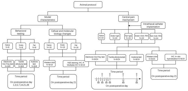
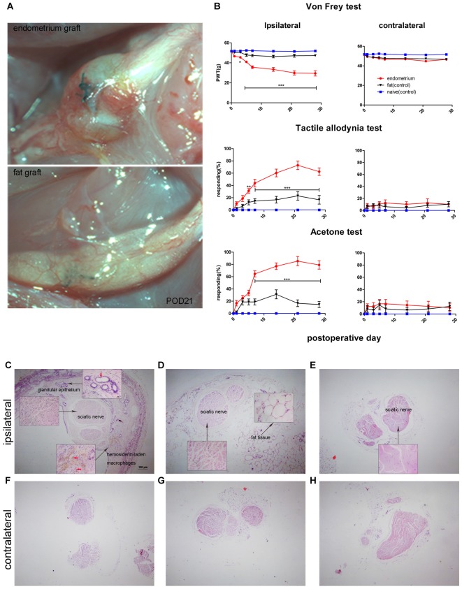
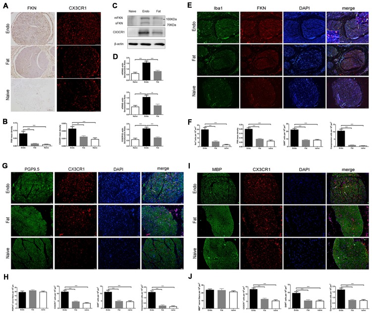
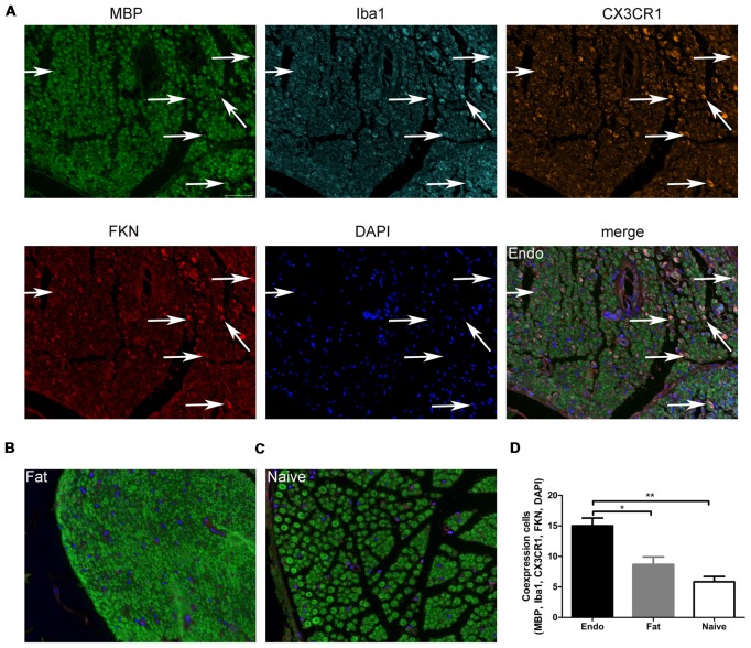
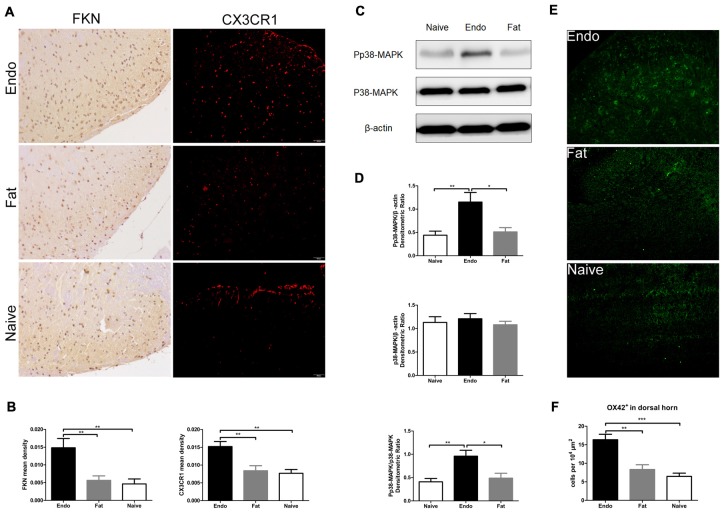
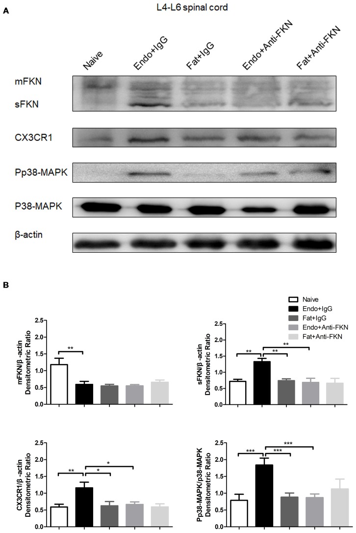
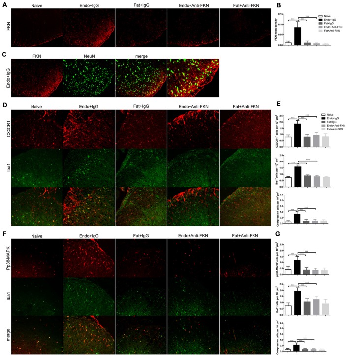
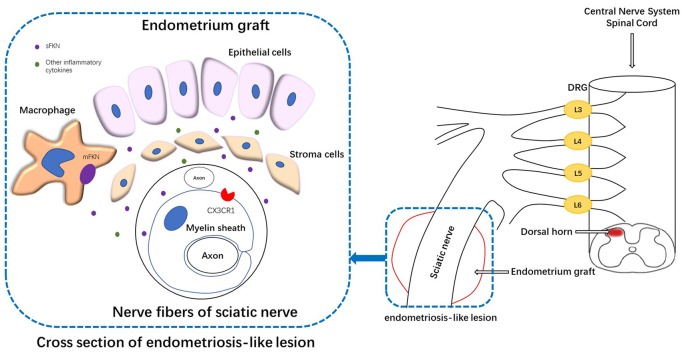
Pain is the most severe and common symptom of endometriosis. Its underlying pathogenetic mechanism is poorly understood. Nerve sensitization is a particular research challenge, due to the limitations of general endometriosis models and sampling nerve tissue from patients. The chemokine fractalkine (FKN) has been demonstrated to play a key role in various forms of neuropathic pain, while its role in endometriotic pain is unknown. Our study was designed to explore the function of FKN in the development and maintenance of peripheral hyperalgesia and central sensitization in endometriosis using a novel endometriosis animal model developed in our laboratory. After modeling, behavioral tests were carried out and the optimal time for molecular changes was obtained. We extracted ectopic tissues and L4-6 spinal cords to detect peripheral and central roles for FKN, respectively. To assess morphologic characteristics of endometriosis-like lesions-as well as expression and location of FKN/CX3CR1-we performed H&E staining, immunostaining, and western blotting analyses. Furthermore, inhibition of FKN expression in the spinal cord was achieved by intrathecal administration of an FKN-neutralizing antibody to demonstrate its function. Our results showed that implanted autologous uterine tissue around the sciatic nerve induced endometriosis-like lesions and produced mechanical hyperalgesia and allodynia. FKN was highly expressed on macrophages, whereas its receptor CX3CR1 was overexpressed in the myelin sheath of sciatic nerve fibers. Overexpressed FKN was also observed in neurons. CX3CR1/pp38-MAPK was upregulated in activated microglia in the spinal dorsal horn. Intrathecal administration of FKN-neutralizing antibody not only reversed the established mechanical hyperalgesia and allodynia, but also inhibited the expression of CX3CR1/pp38-MAPK in activated microglia, which was essential for the persistence of central sensitization. We concluded that the FKN/CX3CR1 signaling pathway might be one of the mechanisms of peripheral hyperalgesia in endometriosis, which requires further studies. Spinal FKN is important for the development and maintenance of central sensitization in endometriosis, and it may further serve as a novel therapeutic target to relieve persistent pain associated with endometriosis.

Keywords: central sensitization; endometriosis; fractalkine; inflammation; microglia; neuropathic pain; peripheral hyperalgesia.


Figures

Figure 1
Figure 1
Flow diagram of the experiment. IHC, immunohistochemistry; IF, immunofluorescence.
Figure 2
Figure 2
Rat sciatic endometriosis model. (A) Uterine grafts formed cysts around the sciatic nerve as visualized on postoperative day 21. (B) Behavioral changes in rats with sciatic endometriosis. Presurgical baseline values (mean of two test points) are presented as post-operative day (POD) 0. Mechanical hyperalgesia was determined by electronic von Frey filament. PWT, paw withdrawal threshold. Tactile allodynia and cold allodynia (acetone test) were measured by the percentage of withdrawal responses to a fine cotton wisp or a drop of acetone. In the uterine graft group, ipsilateral mechanical sensitivity and tactile allodynia were significantly different from the fat control group on all post-surgical days measured except for POD1. The ipsilateral cold allodynia response in the uterine graft group was significantly higher than the fat control group from POD7 to POD28. The mechanical sensitivity and allodynia of the contralateral hind paw showed no significant difference for any group. Two-way repeated-measures analysis of variance (ANOVA) was used for analysis *p < 0.05; **p < 0.01; ***p < 0.001. N = 8 rats per group. (C) H&E staining shows the ipsilateral sciatic nerve surrounded by endometrium, which exhibits typical glandular epithelium (vertical red arrow) and hemosiderin-laden macrophages (horizontal red arrows). Extensive inflammatory cells such as macrophages infiltrated into the nerve, and ectopic endometrium can bevisualized. (D) H&E staining shows the histologic structures in fat tissue with fat vacuoles. Fewer inflammatory cells can be seen in nervous and fat tissue. (E) Histologic features of the untreated sciatic nerve in naïve rats. Fewer inflammatory cells were observed. (F) Histologic features of the contralateral sciatic nerve with fewer inflammatory cells observed in the endometrial graft group. (G) Histologic features of the contralateral sciatic nerve in the fat graft group with fewer inflammatory cells observed. (H) Histologic features of the contralateral sciatic nerve in naïve rats without inflammatory cells observed. (C–H) 40×; insets, 400×. Scale bar for (C–H), 100 μm; scale bar for insets, 20 μm. N = 8 rats per group.
Figure 3
Figure 3
Expressions of fractalkine (FKN) and its receptor CX3CR1 in graft tissue. (A) Immunohistochemical staining for FKN and CX3CR1 in the sciatic nerve of graft tissue. (B) We analyzed FKN and CX3CR1 expression in the sciatic nerve of graft tissue by 1-way ANOVA. (C) Western blotting analysis showed protein levels of membrane-bound FKN, sFKN, and CX3CR1 in graft tissue. (D) Quantification of membrane-bound FKN, sFKN, and CX3CR1 bands in graft tissue, and analysis with 1-way ANOVA. (E) Immunofluorescence staining showed that FKN (red) was mainly expressed in macrophages labeled with Iba1 (green) in graft tissue. (F) The fluorescent images of FKN were statistically graphed for mean density, macrophage-positive cells, DAPI-positive cells, and co-expression in cells under 1-way ANOVA. (G) Immunofluorescent images show that CX3CR1 (red) was highly expressed in nerve fibers as indicated by PGP9.5 (green). The white arrows indicate the co-expression of CX3CR1 and PGP9.5 in cells. (H) Quantitative analysis of CX3CR1-positive cells, PGP9.5-positive cells, DAPI-positive cells, and co-expression in cells using 1-way ANOVA. (I) CX3CR1 was highly expressed on the myelin sheath when co-stained with MBP (green) using immunohistochemical staining. The white arrows indicate the co-expression of CX3CR1 and MBP in cells. (J) Quantitative analysis of CX3CR1-positive cells, MBP-positive cells, DAPI-positive cells, and co-expression in cells using 1-way ANOVA. N = 8 rats per group. **p < 0.01, ***p < 0.001. Arrows show co-expression. Scale bar for (A,E,G,I), 100 μm. Insets, 5 μm.
Figure 4
Figure 4
Myelin sheath, macrophages, and CX3CR1 and FKN expression in sciatic nerve. (A) Immunochemical staining for MBP, Iba1, CX3CR1, FKN, DAPI and the co-expression of these targets in the sciatic nerve of Endo group. (B) MBP, Iba1, CX3CR1, FKN, and DAPI co-expression in the sciatic nerve with fat graft group. (C) MBP, Iba1, CX3CR1, FKN, and DAPI co-expression in the sciatic nerve with Naïve group. (D) The number of positive cells co-expressing MBP, Iba1, CX3CR1, FKN, and DAPI was analyzed by 1-way ANOVA. N = 8 rats per group. *p < 0.05; **p < 0.01. Arrows show co-expression. Scale bar for (A–C), 25 μm.
Figure 5
Figure 5
Increased expression of FKN/CX3CR1, phosphorylated p38-MAPK, and the number of microglia in the spinal cord of rats with sciatic endometriosis. (A) Immunohistochemical staining for FKN and CX3CR1 in the dorsal horn of L4–6 spinal cord. (B) FKN and CX3CR1 expression in the dorsal horn of L4–6 spinal cord was analyzed by 1-way ANOVA. (C) Western blotting experiments showing the protein levels of p38-MAPK phosphorylation in the L4–L6 spinal cord. (D) Quantification of p38-MAPK and pp38-MAPK bands in the L4–L6 spinal cord. (E) The total number of microglia in the dorsal horn was determined by counting OX42-positive cells. (F) Quantitative analysis of OX42-positive cells in the dorsal horn. One-way ANOVA was used for analysis. N = 8 rats per group. *p < 0.05; **p < 0.01; ***p < 0.001. Scale bar for (A), 100 μm.
Figure 6
Figure 6
Enhanced mechanical hypersensitivity induced by endometriosis was reversed by intrathecal injection of FKN-neutralizing antibody. (A) Mechanical hyperalgesia, tactile allodynia, and cold allodynia were alleviated by FKN-neutralizing antibody. from POD7. (B) Endometriosis-associated pain behavior was not alleviated on POD3, but was reversed on POD7, 14, and 21. Two-way repeated-measures ANOVA was used for analysis. N = 8 rats per group. *p < 0.05; **p < 0.01; ***p < 0.001. *Represents the Endo + IgG group vs. Endo + Anti-FKN group. +p < 0.05; +++p < 0.001, +represents the Endo + IgG group vs. Fat + IgG group. #p < 0.05; #represents Fat + Anti-FKN group vs. Endo + Anti-FKN group.
Figure 7
Figure 7
The expression of membrane-bound FKN, sFKN, CX3CR1, and pp38-MAPK in the spinal cord was significantly decreased by intrathecal administration of anti-FKN antibody on POD 21. (A) Western blot images showing protein levels of membrane-bound FKN, sFKN, CX3CR1, and pp38-MAPK in L4–6 spinal cord. (B) Quantification of membrane-bound FKN, sFKN, CX3CR1, and pp38-MAPK bands in the L4-L6 spinal cord. One-way ANOVA was used for analysis. N = 8 rats per group. *p < 0.05; **p < 0.01; ***p < 0.001.
Figure 8
Figure 8
Effects of intrathecal administration of FKN-neutralizing antibody on spinal expression of FKN, CX3CR1, and pp38-MAPK as demonstrated by immunofluorescence staining. (A) Immunofluorescence staining showed expression of FKN in the dorsal horn. Intrathecal administration of FKN-neutralizing antibody markedly inhibited the expression of FKN. (B) Quantitative analysis of FKN mean density in the dorsal horn. (C) FKN (red) was co-expressed (yellow) with NeuN (green). (D) CX3CR1 expression in microglia was decreased in theEndo + Anti-FKN group. The number of activated microglia as determined by Iba1-positive cells was decreased by FKN-neutralizing antibody. (E) Quantitative analysis of CX3CR1-positive cells, Iba1-positive cells, and co-expression of CX3CR1 and Iba1 in dorsal horn cells. (F) Decreased expression of pp38-MAPK in microglia. (G) Quantitative analysis of pp38-MAPK-positive cells, Iba1-positive cells, and positive co-expression of pp38-MAPK and Iba1 in dorsal horn cells. One-way ANOVA was used for analysis. N = 8 rats per group. ***p < 0.001, *represents the Endo + IgG group vs. another group.
Figure 9
Figure 9
Schematic representation of the mechanisms underlying FKN release/action in endometriosis-like lesions. The endometriotic lesions contained a large number of inflammatory cells, including macrophages. The membrane-bound FKN expressed on macrophages may liberate sFKN, binding to its receptor CX3CR1, which is expressed on the myelin sheath of nerve fibers. FKN/CX3CR1 interaction represents a key regulatory mechanism for peripheral hyperalgesia. Nociceptive neuronal sensitization caused by FKN signaling in peripheral lesion reaches the spinal dorsal horn and traverses the DRG, inducing the expression of FKN/CX3CR1 and microglial activation within the dorsal horn; and ultimately develops into central sensitization. DRG, dorsal root ganglion.

Similar articles

Cited by

References

    1. Ahn S. H., Edwards A. K., Singh S. S., Young S. L., Lessey B. A., Tayade C. (2015). IL-17A contributes to the pathogenesis of endometriosis by triggering proinflammatory cytokines and angiogenic growth factors. J. Immunol. 195, 2591–2600. 10.4049/jimmunol.1501138 - DOI - PMC - PubMed
    1. Arnold J., Barcena De Arellano M. L., Ruster C., Vercellino G. F., Chiantera V., Schneider A., et al. . (2012). Imbalance between sympathetic and sensory innervation in peritoneal endometriosis. Brain Behav. Immun. 26, 132–141. 10.1016/j.bbi.2011.08.004 - DOI - PubMed
    1. Arosh J. A., Lee J., Balasubbramanian D., Stanley J. A., Long C. R., Meagher M. W., et al. . (2015). Molecular and preclinical basis to inhibit PGE2 receptors EP2 and EP4 as a novel nonsteroidal therapy for endometriosis. Proc. Natl. Acad. Sci. U S A 112, 9716–9721. 10.1073/pnas.1507931112 - DOI - PMC - PubMed
    1. Asante A., Taylor R. N. (2011). Endometriosis: the role of neuroangiogenesis. Annu. Rev. Physiol. 73, 163–182. 10.1146/annurev-physiol-012110-142158 - DOI - PubMed
    1. As-Sanie S., Harris R. E., Harte S. E., Tu F. F., Neshewat G., Clauw D. J. (2013). Increased pressure pain sensitivity in women with chronic pelvic pain. Obstet. Gynecol. 122, 1047–1055. 10.1097/AOG.0b013e3182a7e1f5 - DOI - PMC - PubMed