Non-linearities in the Rod and Cone Photoreceptor Inputs to the Afferent Pupil Light Response
- PMID: 30622511
- PMCID: PMC6308191
- DOI: 10.3389/fneur.2018.01140
Non-linearities in the Rod and Cone Photoreceptor Inputs to the Afferent Pupil Light Response
Abstract
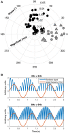
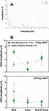
Purpose: To assess the nature and extent of non-linear processes in pupil responses using rod- and cone-isolating visual beat stimuli. Methods: A four-primary photostimulating method based on the principle of silent substitution was implemented to generate rod or cone isolating and combined sinusoidal stimuli at a single component frequency (1, 4, 5, 8, or 9 Hz) or a 1 Hz beat frequency (frequency pairs: 4 + 5, 8 + 9 Hz). The component frequencies were chosen to minimize the melanopsin photoresponse of intrinsically photosensitive retinal ganglion cells (ipRGCs) such that the pupil response was primarily driven by outer retinal photoreceptor inputs. Full-field (Ganzfeld) pupil responses and electroretinograms (ERGs) were recorded to the same stimuli at two mesopic light levels (-0.9 and 0 log cd/m2). Fourier analysis was used to derive the amplitudes and phases of the pupil and ERG responses. Results: For the beat frequency condition, when modulation was restricted to the same photoreceptor type at the higher mesopic level (0 log cd/m2), there was a pronounced pupil response to the 1 Hz beat frequency with the 4 + 5 Hz frequency pair and rare beat responses for the 8 + 9 Hz frequency pair. At the lower mesopic level there were few and inconsistent beat responses. When one component modulated the rod excitation and the other component modulated the cone excitation, responses to the beat frequency were rare and lower than the 1 Hz component frequency condition responses. These results were confirmed by ERG recordings. Conclusions: There is non-linearity in both the pupil response and electroretinogram to rod and cone inputs at mesopic light levels. The presence of a beat response for modulation components restricted to a single photoreceptor type, but not for components with cross-photoreceptor types, indicates that the location of a non-linear process in the pupil pathway occurs at a retinal site earlier than where the rod and cone signals are combined, that is, at the photoreceptor level.
Keywords: ERG analysis; beats; mesopic light level; non-linearity; photoreceptors cells; pupil; retina.
Figures


Similar articles
-
Assessing rod, cone, and melanopsin contributions to human pupil flicker responses.Invest Ophthalmol Vis Sci. 2014 Feb 4;55(2):719-27. doi: 10.1167/iovs.13-13252. Invest Ophthalmol Vis Sci. 2014. PMID: 24408974 Free PMC article.
-
Melanopsin and Cone Photoreceptor Inputs to the Afferent Pupil Light Response.Front Neurol. 2019 May 22;10:529. doi: 10.3389/fneur.2019.00529. eCollection 2019. Front Neurol. 2019. PMID: 31191431 Free PMC article.
-
The Roles of Rods, Cones, and Melanopsin in Photoresponses of M4 Intrinsically Photosensitive Retinal Ganglion Cells (ipRGCs) and Optokinetic Visual Behavior.Front Cell Neurosci. 2018 Jul 12;12:203. doi: 10.3389/fncel.2018.00203. eCollection 2018. Front Cell Neurosci. 2018. PMID: 30050414 Free PMC article.
-
The assessment of L- and M-cone specific electroretinographical signals in the normal and abnormal human retina.Prog Retin Eye Res. 2003 Sep;22(5):579-605. doi: 10.1016/s1350-9462(03)00049-1. Prog Retin Eye Res. 2003. PMID: 12892643 Review.
-
[Pupil and melanopsin photoreception].Nippon Ganka Gakkai Zasshi. 2013 Mar;117(3):246-68; discussion 269. Nippon Ganka Gakkai Zasshi. 2013. PMID: 23631256 Review. Japanese.
Cited by
-
Photoreceptor inputs to pupil control.J Vis. 2019 Aug 1;19(9):5. doi: 10.1167/19.9.5. J Vis. 2019. PMID: 31415056 Free PMC article.
-
Deep learning-based pupil model predicts time and spectral dependent light responses.Sci Rep. 2021 Jan 12;11(1):841. doi: 10.1038/s41598-020-79908-5. Sci Rep. 2021. PMID: 33436693 Free PMC article.
-
PySilSub: An open-source Python toolbox for implementing the method of silent substitution in vision and nonvisual photoreception research.J Vis. 2023 Jul 3;23(7):10. doi: 10.1167/jov.23.7.10. J Vis. 2023. PMID: 37450287 Free PMC article.
-
Spatial Cluster Patterns of Retinal Sensitivity Loss in Intermediate Age-Related Macular Degeneration Features.Transl Vis Sci Technol. 2023 Sep 1;12(9):6. doi: 10.1167/tvst.12.9.6. Transl Vis Sci Technol. 2023. PMID: 37676679 Free PMC article.
-
Prediction accuracy of L- and M-cone based human pupil light models.Sci Rep. 2020 Jul 3;10(1):10988. doi: 10.1038/s41598-020-67593-3. Sci Rep. 2020. PMID: 32620793 Free PMC article.
References
Grants and funding
LinkOut - more resources
Full Text Sources

