. 2018 Dec 21:9:3043.
doi: 10.3389/fimmu.2018.03043. eCollection 2018.

Type 1 Conventional CD103+ Dendritic Cells Control Effector CD8+ T Cell Migration, Survival, and Memory Responses During Influenza Infection

Affiliations

Type 1 Conventional CD103+ Dendritic Cells Control Effector CD8+ T Cell Migration, Survival, and Memory Responses During Influenza Infection

See Liang Ng et al. Front Immunol. .

Abstract

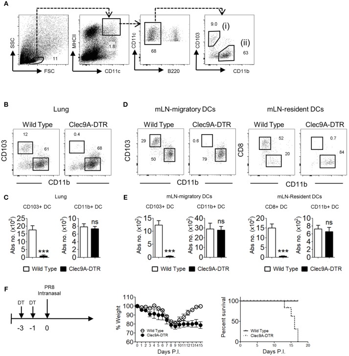
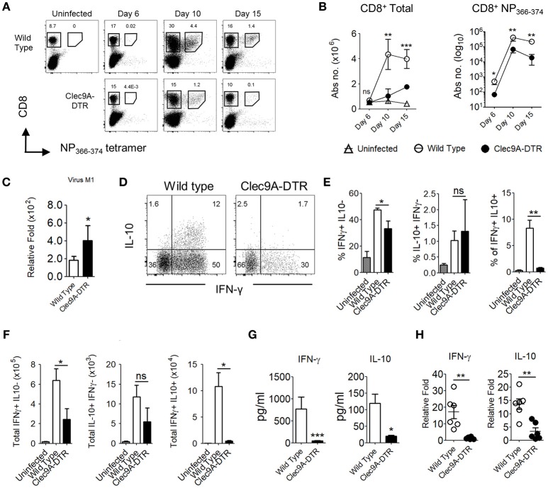
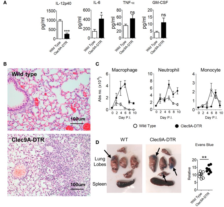
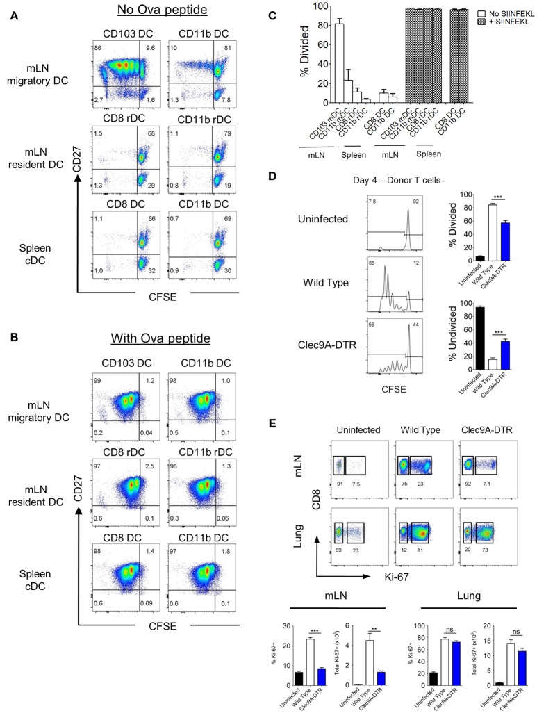
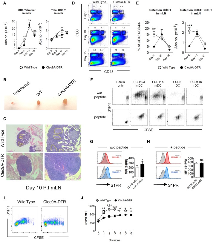
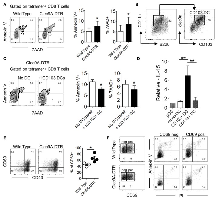
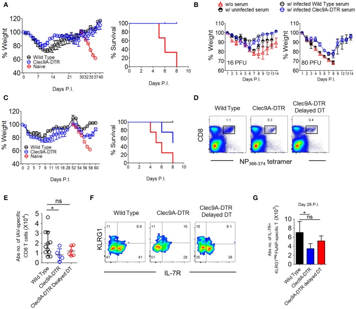
Type 1 conventional CD103+ dendritic cells (cDC1) contribute significantly to the cytotoxic T lymphocyte (CTL) response during influenza virus infection; however, the mechanisms by which cDC1s promote CTL recruitment and viral clearance are unclear. We demonstrate that cDC1 ablation leads to a deficient influenza-specific primary CD8+ T cell response alongside severe pulmonary inflammation, intensifying susceptibility to infection. The diminished pulmonary CTL population is not only a consequence of reduced priming in the lymph node (LN), but also of dysregulated CD8+ T cell egression from the LN and reduced CD8+ T cell viability in the lungs. cDC1s promote S1PR expression on CTLs, a key chemokine receptor facilitating CTL LN egress, and express high levels of the T cell survival cytokine, IL-15, to support CTL viability at the site of infection. Moreover, cDC1 ablation leads to severe impairment of CD8+ T cell memory recall and cross-reactive protection, suggesting that cDC1 are not only involved in primary T cell activation, but also in supporting the development of effective memory CD8+ T cell precursors. Our findings demonstrate a previously unappreciated and multifaceted role of CD103+ DCs in controlling pulmonary T cell-mediated immune responses.

Keywords: CD103; CD8+ T cell; Clec9A; dendritic cell; inflammation; influenza; migration; survival.


Figures

Figure 1
Figure 1
Clec9A-DTR mice succumb to a sub-lethal influenza infection. (A) Gating strategy for migratory CD103+ cDC1 in the lung, defined as MHCIIhiCD11chiB220CD11bCD103+ (B) Representative ablation profile of migratory CD103+ cDC1s in the lung of Clec9A-DTR mice after 2 consecutive days of DT administration. (C) Absolute numbers of migratory cDCs in the lung of DT-treated wild type and Clec9A-DTR mice. (D) Representative ablation profile of migratory CD103+ cDC1s and resident CD8+ cDC1s in the mLN of Clec9A-DTR mice after two consecutive days of DT administration. (E) Absolute numbers of migratory and resident cDCs in the mLN of DT-treated wild type and Clec9A-DTR mice. (F) Representative weight loss (left) and survival (right) curve of wild type and Clec9A-DTR mice after 16 PFU of PR8-H1N1 infection. Data are shown as mean ± SEM. ***p < 0.001. Data represent two (C,E) (n = 3–5) or three (F) (n = 4) independent experiments.
Figure 2
Figure 2
Impaired effector CD8+ T cell responses in the absence of pulmonary CD103+ cDC1s. (A) NP366−374-specific CD8+ T cells in the lungs of uninfected, infected wild type, and Clec9A-DTR mice (day 6, 10, and 15 post infection). (B) Kinetics of total CD8+ T cells and NP366−374-specific CD8+ T cells in the lungs of uninfected, infected wild type, and Clec9A-DTR mice. Absolute numbers are shown. (C) Lung virus load was measured by relative quantification of M1 viral protein in infected wild type and Clec9A-DTR mice on day 10 post infection. (D–F) Lung cells were harvested from uninfected, infected wild type, and Clec9A-DTR mice on day 10 post infection and stimulated with PMA/Ionomycin for 3 h followed by Brefeldin A incubation for an additional 3 h. Intracellular IFN-γ and IL-10 staining profiles (d) of pulmonary CD8+ T cells, frequency (E) and total numbers (F) of IFN-γ-producing, IL-10-producing, and IFN-γ/IL-10 double-producing CD8+ T cells in the lungs. (G) IFN-γ and IL-10 BAL levels as measured by sandwich ELISA (H) Relative quantification of IFN-γ and IL-10 transcripts. Data are shown as mean ± SEM. *p < 0.05. **p < 0.01. ***p < 0.001. Data represent two (B–F) (n = 3–5) independent experiments.
Figure 3
Figure 3
Enhanced pulmonary inflammation and severe lung damage in IAV-infected Clec9A-DTR mice. (A) IL-12p40, IL-6, TNF-α, and GM-CSF BAL levels measured by sandwich ELISA (day 10 post infection). (B) Representative H&E histology images of lungs from day 10-infected wild type and Clec9A-DTR mice. Magnification 80×. (C) Total number of macrophages, neutrophils, and monocytes in the lungs on day 2, 4, 6, 8, and 10 post infection. (D) Images of lungs from day 10-infected wild type and Clec9A-DTR mice 30 min after administration of Evans blue dye (left and middle). Relative quantification calculated from absorbance readings at OD610 (right). Black arrows indicate Evans blue dense regions. Data are shown as mean ± SEM. *p < 0.05. **p < 0.01. ***p < 0.001. Data represent two (A–D) (n = 3–5) independent experiments.
Figure 4
Figure 4
CD103+, but not CD8+ cDC1s, cross -present virus antigen in the mLN. (A,B) Spleen and mLN DCs were sorted from wild type mice infected with recombinant OT1-PR8 virus for 5 days. Sorted DCs were individually co-cultured with CFSE-labeled OT1-CD8+ T cells. CFSE dilution profiles of OT1-CD8+ T cells after 4 days co-culture with respective DCs without (A) or with (B) the addition of exogenous ova peptide OVA257−264. (C) Proportion of OT1-CD8+ T cells that have undergone at least one cell division. (D) CFSE dilution profiles of donor OT1-CD8+ T cells (Ly5.1+ and CD8+) in the mLN (left). Wild type and Clec9A-DTR recipients were infected with recombinant OT1-PR8 virus 3 h before donor OT1-CD8+ T cells transfer (2 × 10⁶), and mLNs were harvested 4 days after infection. Proportion of OT1-CD8+ T cells which has either undergone at least one cell division (top right) or no division (bottom right). (E) Lungs and mLN were harvested from mice infected with PR8 virus for 10 days. Profiles of intracellular staining for Ki-67 by CD8+ T cells in the mLN and lungs. Total number and frequency of Ki-67+CD8+ T cells in both mLN and lungs. mDC, migratory DC. rDC, resident DC. Data are shown as mean ± SEM. **p < 0.01, ***p < 0.001. Data represent two (C–E) (n = 3–5) independent experiments.
Figure 5
Figure 5
CD103+ cDC1s support the LN egression of IAV-specific CD8+ T cells. (A) Total number of NP366−374-specific CD8+ T cells (left) and bulk CD8+ T cells (right) in the mLN of wild type and Clec9A-DTR mice on day 3, 6, 10, and 15 of infection. (B) Representative picture of posterior mLN harvested from uninfected mice and infected Clec9A-DTR and wild type mice after 10 days of infection. (C) Representative H&E histology images of posterior mLN harvested from day 10-infected Clec9A-DTR and wild type mice. (D) Representative flow cytometry profiles of CD8 and CD43 stained cells obtained from mLN harvested on day 6, 10, and 15 of infection. (E) Frequency of CD43+/CD43 CD8+ T cells in the mLN (left). Total number of CD43+ CD8+ T cells in the mLN (right) on day 6, 10, and 15 of infection. (F) Flow cytometry profiles of OT1+CD8+ T cells co-cultured with sorted DCs for 3 days with or without exogenous SIINFEKL peptide. Cells are gated on Ly5.1+, CD8+, and CD3+. (G) MFI of S1PR by OT1+CD8+ T cells that were co-cultured without DC (gray filled), with migratory CD103+ cDC1s (blue), or with migratory CD11b+ cDC2s (red) in the absence of exogenous SIINFEKL peptide. (H) MFI of S1PR by OT1+CD8+ T cells that were co-cultured without DC (gray filled), with migratory CD103+ cDC1s (blue), or with migratory CD11b+ cDC2s (red) in the presence of exogenous SIINFEKL peptide. (I) CFSE dilution profiles of donor OT1-CD8+ T cells in the mLN. Wild type and Clec9A-DTR recipients were infected with recombinant OVA-PR8 virus 3 h before donor OT1-CD8+ T cells transfer (2 × 10⁶), and mLNs were harvested 4 days after infection. (J) MFI of S1PR expression by donor OT1-CD8+ T cells in each cell division. Data are shown as mean ± SEM. *p < 0.05. **p < 0.01. ***p < 0.001. Data represent two (A,E,G–J) (n = 3–5) independent experiments.
Figure 6
Figure 6
CD103+ cDC1s alter effector CD8+ T cell survival in IAV-infected lungs. (A) Annexin V and 7AAD staining profile of NP366−374-specific CD8+ T cells in the lung of wild type and Clec9A-DTR mice on day 10 of infection (left). Frequency of Annexin V+/7AAD+ NP366−374-specific CD8+ T cells (right). (B) Characterization of BM-differentiated CD103+ cDC1s (iCD103+ cDC1s) after 16 days in culture supplemented with GM-CSF and Flt3L. (C) Annexin V and 7AAD staining profile of NP366−374-specific CD8+ T cells in the lung of Clec9A-DTR recipient mice with or without adoptive transfer of iCD103+ cDC1s on day 11 infection (left). Frequency of Annexin V+/7AAD+ NP366−374-specific CD8+ T cells (right). (D) Relative mRNA expression of IL-15 by plasmacytoid DCs, monocyte-derived DCs, migratory CD103+ cDC1s, and migratory CD11b+ cDC2s from infected wild type mice on day 10 infection. (E) Representative CD69 and CD43 staining profile of NP366−374-specific CD8+ T cells in the lung of wild type and Clec9A-DTR mice on day 10 of infection (left) and calculated frequencies of CD69+ NP366−374-specific CD8+ T cells (right). (F) Representative CD69+ and CD69 gates on NP366−374-specific CD8+ T cells in the lung of wild type and Clec9A-DTR mice on day 10 infection (left) and representative surface expression profiles of Annexin V+ and PI+ by CD69+ and CD69 populations. Data are shown as mean ± SEM. *p < 0.05. **p < 0.01. Data represent two (A,C–F) (n = 3–5) independent experiments.
Figure 7
Figure 7
CD103+ cDC1s are essential for cross-reactive protection against serotypically distinct virus subtypes. (A) Wild type and Clec9A-DTR mice were infected with 3 PFU x-31 virus and subsequently infected with 600 PFU x-31 virus after 30 days. Naïve mice were infected with 600 PFU x-31 virus on day 30 only. Weight loss curve (left) and survival curve (right) for mice after 600 PFU x-31 virus infection. (B) Sera were harvested from naïve, infected wild type, and Clec9A-DTR mice. Wild type recipient mice were administered with 50 μl of collected serum 2 days before infection with 16 or 80 PFU PR8 virus. Wild type mice without serum transfer were used as controls. Weight loss curve of recipients which received 16 PFU (left) and 80 PFU (right) PR8 virus. (C) Wild type and Clec9A-DTR mice were infected with 3 PFU x-31 virus and subsequently infected with 600 PFU PR8 virus after 28 days. Naïve mice were infected with 600 PFU PR8 virus on day 28 only. Weight loss curve (left) and survival curve for mice after 600 PFU PR8 virus infection (right). (D,E) Representative flow cytometry plots and absolute numbers of NP366−374-specific CD8+ T cells in the lungs of wild type, Clec9A-DTR and Clec9A-DTR delayed DT injected mice on day 35 of infection. Clec9A-DTR mice were injected with DT from day−5 to day 14 of infection. Clec9A-DTR delayed DT mice were injected with DT from day 21 to day 35 of infection. (F) Representative flow cytometry staining of KLRG1 and IL7R expression by NP366−374-specific CD8+ T cells (day 35 P.I). (G) Absolute number of IL7R+ NP366−374-specific CD8+ T cells in the lungs (day 35 P.I). Data are shown as mean ± SEM. *p < 0.05. Data represent two [(A,B) (n = 3–5) and (C,E,G) (n = 3–4)] independent experiments.

References

    1. GeurtsvanKessel CH, Willart MA, van Rijt LS, Muskens F, Kool M, Baas C, et al. Clearance of influenza virus from the lung depends on migratory langerin+CD11b- but not plasmacytoid dendritic cells. J Exp Med. (2008) 205:1621–34. 10.1084/jem.20071365 - DOI - PMC - PubMed
    1. Ballesteros-Tato A, Leon B, Lund FE, Randall TD. Temporal changes in dendritic cell subsets, cross-priming and costimulation via CD70 control CD8(+) T cell responses to influenza. Nat Immunol. (2010) 11:216–24. 10.1038/ni.1838 - DOI - PMC - PubMed
    1. Legge KL, Braciale TJ. Accelerated migration of respiratory dendritic cells to the regional lymph nodes is limited to the early phase of pulmonary infection. Immunity (2003) 18:265–77. 10.1016/S1074-7613(03)00023-2 - DOI - PubMed
    1. McGill J, Legge KL. Cutting edge: contribution of lung-resident T cell proliferation to the overall magnitude of the antigen-specific CD8 T cell response in the lungs following murine influenza virus infection. J Immunol. (2009) 183:4177–81. 10.4049/jimmunol.0901109 - DOI - PMC - PubMed
    1. Ray SJ, Franki SN, Pierce RH, Dimitrova S, Koteliansky V, Sprague AG, et al. . The collagen binding alpha1beta1 integrin VLA-1 regulates CD8 T cell-mediated immune protection against heterologous influenza infection. Immunity (2004) 20:167–79. 10.1016/S1074-7613(04)00021-4 - DOI - PubMed

Publication types

MeSH terms