. 2018 Dec 3:2018:7283703.
doi: 10.1155/2018/7283703. eCollection 2018.

Mesenchymal Stem Cells Ameliorate Hepatic Ischemia/Reperfusion Injury via Inhibition of Neutrophil Recruitment

Affiliations

Mesenchymal Stem Cells Ameliorate Hepatic Ischemia/Reperfusion Injury via Inhibition of Neutrophil Recruitment

Shihui Li et al. J Immunol Res. .

Abstract

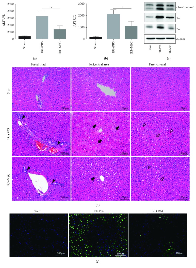
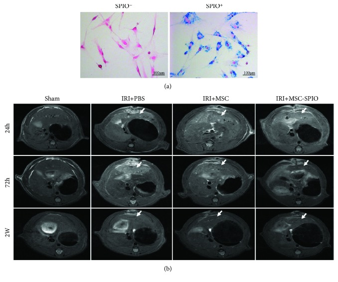
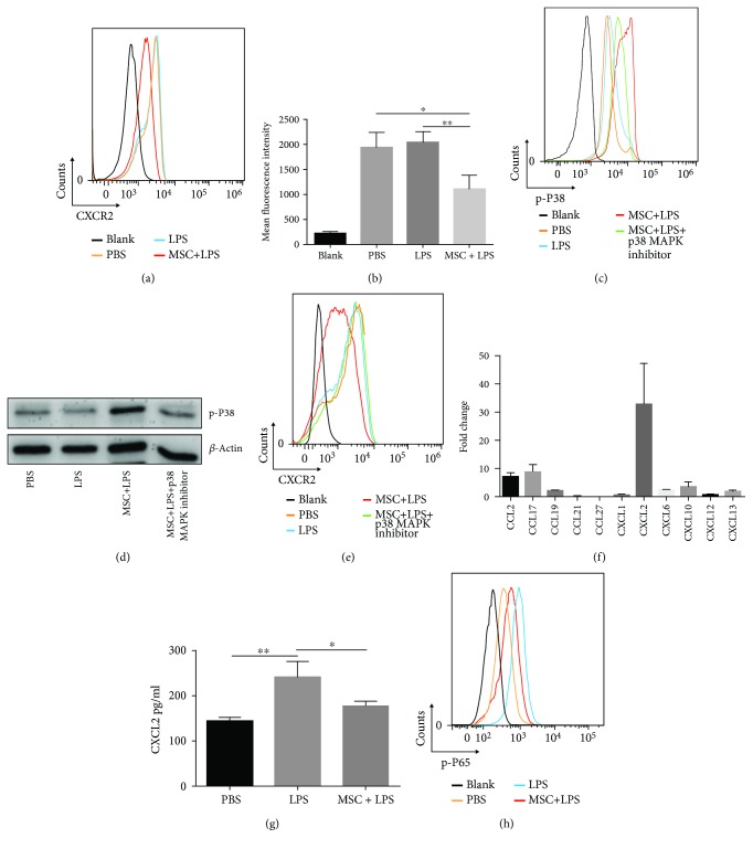
Ischemia/reperfusion injury (IRI) remains a major problem in organ transplantation, which represents the main cause of graft dysfunction posttransplantation. Hepatic IRI is characterized by an excessive inflammatory response within the liver. Mesenchymal stem cells (MSCs) have been shown to be immunomodulatory cells and have the therapeutic action on IRI in several organs. However, the mechanism of regulatory effect of MSCs on IRI remains unclear. In the present study, we examined the impact of MSCs on hepatic inflammatory response such as neutrophil influx and liver damage in a rat model of 70% hepatic IRI. Treatment with MSCs protected rat against hepatic IRI, with significantly decreased serum levels of liver enzymes, attenuated hepatic neutrophil infiltration, reduced expression of apoptosis-associated proteins, and ameliorated liver pathological injury. MSCs also significantly enhanced the intracellular activation of p38 MAPK phosphorylation, which led to decreased expression of CXCR2 on the surface of neutrophils. In addition, MSCs significantly diminished neutrophil chemoattractant CXCL2 production by inhibiting NF-κB p65 phosphorylation in macrophages. These results demonstrate that MSCs significantly ameliorate hepatic IRI predominantly through its inhibitory effect on hepatic neutrophil migration and infiltration.


Figures

Figure 1
Figure 1
Characterization of rat bone marrow derived-MSCs. (a) Immunophenotype of rat MSCs. Cells were harvested at passage 3, labeled with the antibodies specific for the indicated surface antigens or negative controls, and analyzed by flow cytometry. The numbers in the panels represent the mean fluorescence intensity of the cells expressing each marker. (b) Rat MSCs at passage 3 that were induced to differentiate into adipocytes and osteoblasts. Cells stain positive for oil with oil red staining and for calcium with alizarin red solution, respectively. Original magnification, ×200.
Figure 2
Figure 2
Rat MSCs reduced the release of liver enzymes and improved the histopathologic changes of livers in IRI. Male SD rats were randomized into sham, IRI + PBS, and IRI + MSC groups. Samples of each group were collected 12 h postreperfusion. (a, b) As markers for hepatic injury, serum levels of ALT and AST were determined. (c) The expression levels of various proapoptotic proteins including Cleaved caspase-3, Bad, and Fas in the I/R liver lobes were determined by Western blot analysis. (d) Representative images of hematoxylin and eosin-stained sections of liver tissues were shown. Inflammatory cell accumulation (solid black triangles), hepatocellular swelling (solid black arrows), necrosis (hollow arrows), and acidophilic degeneration (hollow triangles) were observed. (e) TUNEL staining of apoptosis cells (bright green) in liver tissues. Data are mean ± SD; p < 0.05. Original magnification, ×200.
Figure 3
Figure 3
MSCs affect cytokine expression and reduce neutrophil infiltration. (a, e, f) The relative mRNA levels of IL-2, IL-4, IL-6, IL-10, CXCL2, CD11b, and CD18 genes in I/R lobes were detected by qRT-PCR. Data were normalized to glyceraldehyde-3-phosphate dehydrogenase gene expression. (b) The concentration of IL-2, IL-4, IL-6, IL-10, and CXCL2 in I/R lobes was measured by ELISA. (c) Representative images of immunohistochemistry-stained sections of MPO+ cells of liver tissues were shown. (d) The mean frequency of hepatic MPO+ cells in 10 high-power fields was calculated. Data are mean ± SD. p < 0.05, ∗∗p < 0.01, ∗∗∗p < 0.001. Original magnification, ×200.
Figure 4
Figure 4
MSCs accumulated in injured liver lobes in the model of hepatic IRI. (a) SPIO-labeled MSCs were positive for Prussian blue staining. Blue particles were observed in the cytoplasm of SPIO+ cells. (b) Male SD rats were randomized into sham, IRI + PBS, IRI + MSC, and IRI + MSC-SPIO groups. Representative images of MRI scanning on the T2 sequence of I/R lobes in each group were shown after 24 h, 72 h, and 2 w of reperfusion. The liver lobes experienced IRI were pointed out by white arrows. Original magnification, ×200.
Figure 5
Figure 5
MSCs attenuate neutrophil recruitment via hindering CXCL2/CXCR2 signaling. (a, e) Surface expression of CXCR2 on neutrophils was detected by flow cytometry analysis. (b) The mean fluorescence intensity of CXCR2 on the surface of neutrophils was recorded. (c) The levels of p38 MAPK phosphorylation in neutrophils were detected by flow cytometry analysis, and (d) the consequences were verified by Western blot analysis. (f) The expression levels of mRNA of various chemokines involved in hepatic IRI were analyzed by qRT-PCR. Fold change represents the expression of each chemokine in I/R liver lobes 12 h postreperfusion compared with normal liver. (g) Macrophages were cultured in the presence or absence of MSCs for 12 h. The concentration of CXCL2 in the supernatant was measured by ELISA. (h) The levels of NF-κB p65 phosphorylation in macrophages were detected by flow cytometry analysis. Data are mean ± SD. p < 0.05, ∗∗p < 0.01, ∗∗∗p < 0.001. p-P38: phospho-p38 MAPK; p-P65: phospho-NF-κB p65.

Similar articles

Cited by

References

    1. Kim W. R., Lake J. R., Smith J. M., et al. OPTN/SRTR 2013 annual data report: liver. American Journal of Transplantation. 2015;15(Supplement 2):1–28. doi: 10.1111/ajt.13197. - DOI - PubMed
    1. Serracino-Inglott F., Habib N. A., Mathie R. T. Hepatic ischemia-reperfusion injury. American Journal of Surgery. 2001;181(2):160–166. doi: 10.1016/S0002-9610(00)00573-0. - DOI - PubMed
    1. Busuttil R., Tanaka K. The utility of marginal donors in liver transplantation. Liver Transplantation. 2003;9(7):651–663. doi: 10.1053/jlts.2003.50105. - DOI - PubMed
    1. Saidi R. F., Kenari S. K. H. Liver ischemia/reperfusion injury: an overview. Journal of Investigative Surgery. 2014;27(6):366–379. doi: 10.3109/08941939.2014.932473. - DOI - PubMed
    1. Cannistra M., Ruggiero M., Zullo A., et al. Hepatic ischemia reperfusion injury: a systematic review of literature and the role of current drugs and biomarkers. International Journal of Surgery. 2016;33:S57–S70. doi: 10.1016/j.ijsu.2016.05.050. - DOI - PubMed

Supplementary concepts

LinkOut - more resources