. 2018 Dec 31;3(6):766-778.
doi: 10.1016/j.jacbts.2018.08.006. eCollection 2018 Dec.

Characterization of the Myocardial Inflammatory Response in Acute Stress-Induced (Takotsubo) Cardiomyopathy

Affiliations

Characterization of the Myocardial Inflammatory Response in Acute Stress-Induced (Takotsubo) Cardiomyopathy

Heather M Wilson et al. JACC Basic Transl Sci. .

Abstract

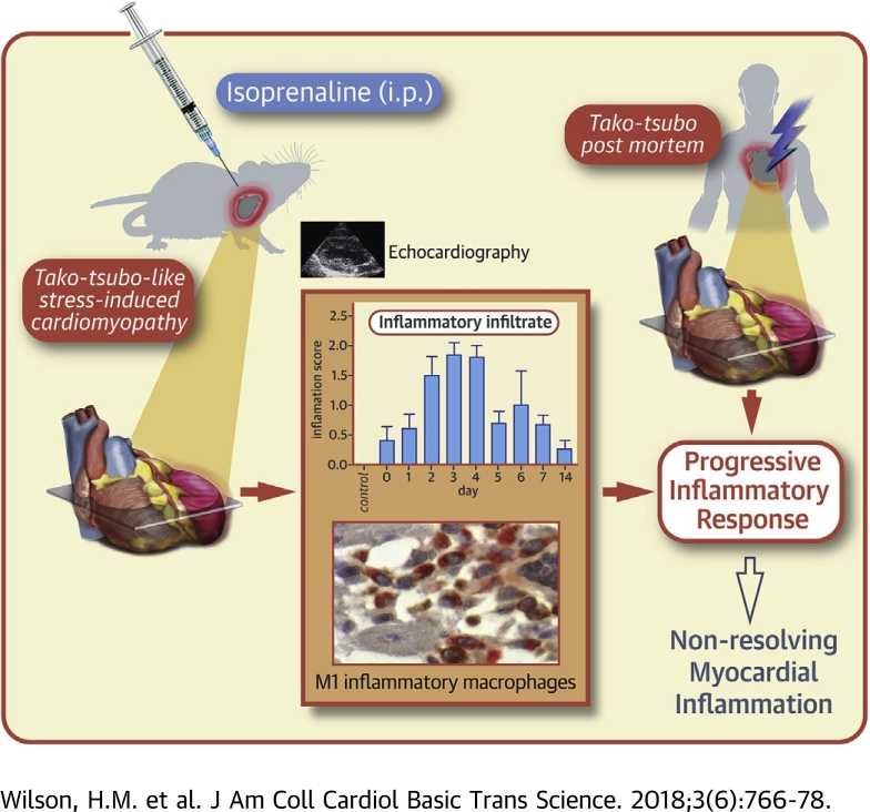
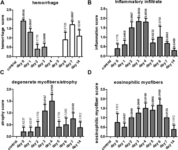
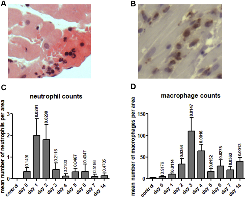
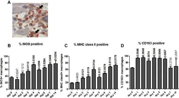
Takotsubo cardiomyopathy is an acute stress-induced heart failure syndrome for which the exact pathogenic mechanisms are unclear, and consequently, no specific treatment exists. In an experimental model of stress-induced takotsubo-like cardiomyopathy, the authors describe the temporal course of a chronic inflammatory response post-induction, with an initial early influx of neutrophils into myocardial tissue followed by macrophages that are typical of a proinflammatory M1 phenotype, and a nonsignificant increase in systemic inflammatory cytokines. Post-mortem myocardium from the more complex clinical takotsubo patients share features of the study's experimental model. These findings suggest modulators of inflammation could be a potential therapeutic option.

Keywords: EF, ejection fraction; IL, interleukin; MHC, major histocompatibility complex; MI, myocardial infarction; TNFα, tumor necrosis factor-alpha; histopathology; inflammation; macrophage; pathophysiology; qPCR, quantitative polymerase chain reaction; takotsubo cardiomyopathy.


Figures

None
Graphical abstract
Figure 1
Figure 1
Histological Characterization of Mid-Heart Sections of Control Rats and From Rats After Induction of Takotsubo Cardiomyopathy Histological characterization of hematoxylin and eosin–stained mid-heart sections of control rats and from rats over the time course (0 to 14 days) after induction of takotsubo cardiomyopathy show a temporal immune/inflammatory cellular infiltrate. Sections of heart tissue were semiquantitatively scored by a consultant histopathologist (K.K.) blinded to the study. Arbitrary scores of 0, 1, and 2 were given for (A) hemorrhage (gray bars) or presence of hemosiderin-containing phagocytic cells (white bars), (B) immune/inflammatory cellular infiltrates, (C) degenerate myofibrils/myofiber atrophy, and (D) eosinophilic myofibers. The number of animals per group were as follows; control n = 7, day 1 n = 6, day 2 n = 6, day 3 n = 7, day 4 n = 6, day 5 n = 7, day 6 n = 6, day 7 n = 5, and day 14 n = 5 animals. Statistical difference in histological scores between control rats and those injected with isoprenaline were calculated with p values denoted above each time point bar. Values in bold indicate statistically significant differences compared with the control group.
Figure 2
Figure 2
Immunohistochemical Analysis Following Induction of Stress-Induced Takotsubo-Like Cardiomyopathy Immunohistochemical analysis demonstrates progressive neutrophil and macrophage infiltration into myocardial tissue (day 0 to 14) following induction of stress-induced takotsubo-like cardiomyopathy. Representative examples of neutrophil (A) and macrophage (CD68-positive cells) (B) staining in mid-region rat myocardial tissue, original magnification ×400. Neutrophil (C) and macrophage (D) cell counts in the mid-region of saline injected (control) and isoprenaline injected rat hearts (day 0 to 14). Values represent mean counts ± SD in 24 fields of view from defined areas as described in the Methods section. The number of animals per group were as follows: control n = 7, day 1 n = 6, day 2 n = 6, day 3 n = 7, day 4 n = 6, day 5 n = 7, day 6 n = 6, day 7 n = 5, and day 14 n = 5 animals. Statistical difference in mean number of neutrophil and macrophage counts between control rats and those injected with isoprenaline were calculated with p values denoted above each time point bar. Values in bold indicate statistically significant differences compared with the control group.
Figure 3
Figure 3
Double Immunostaining of Mid-Heart Sections Showing M1-Activated and M2-Positive Macrophages Double immunostaining of mid-heart sections show a greater predominance of M1-activated macrophages over time with little change in percentage of M2-positive macrophages. Total macrophages were detected by anti-CD68 and liquid permanent red staining (red), whereas macrophage activation markers were identified by the relevant antibodies and DAB staining (brown). Macrophage-rich areas were analyzed as outlined in Methods. Examples of double-stained macrophages are highlighted by arrows in A. Original magnification ×400. Changes in percentages of double-stained macrophages over time for inducible nitric oxide synthase (iNOS) (B), major histocompatibility complex (MHC) class II (C), and CD163 (D). The number of animals per group were as follows; control n = 6, day 1 to 14 n = 5 for each time point. Statistical difference in the percentage iNOS, MHC class II, and CD163-positive macrophages between control rats and those injected with isoprenaline were calculated with p values denoted above each bar. Values in bold indicate statistically significant differences compared with the control group.
Figure 4
Figure 4
Correlations Between the Total Macrophage Counts or the Percentage M1 and M2 Macrophages and Recovery of Cardiac Function Correlations between the total macrophage counts or the percentage M1 and M2 macrophages and recovery of cardiac function as determined by percentage left ventricular ejection fraction (EF). The total number of macrophages (A) or percentage of iNOS (B) and MHC class II–positive (C) M1 macrophages and CD163-positive (D) M2-like macrophages was determined from immunohistochemical analysis and correlated with the percentage ejection fraction before cull. A total of 13 animals were analyzed. The coefficient of determination (R2) and p value were determined by Pearson’s correlation test.
Figure 5
Figure 5
Analysis of Systemic and Myocardial Cytokine Levels in Control Animals and Those With Takotsubo-Like Cardiomyopathy Analysis of systemic and myocardial cytokine levels in control animals and those with Takotsubo-like cardiomyopathy at day 3 (d3) and day 14 (d14) are shown. Serum levels of IL-6 (A) and IL-10 (B) are mean pg/ml levels ± SD; gene expression levels of IL-6 (C) and IL-10 (D). Groups analyzed consisted of control, saline-injected animals (n = 10), isoprenaline-injected animals sacrificed 3 days after injection (n = 9), and isoprenaline-injected animals sacrificed 14 days after injection (n = 5). Values are mean fold change over control animals ± SD. Values above each bar indicate p values compared with control animals. IL = interleukin.
Figure 6
Figure 6
Post-Mortem Myocardial Tissue From Takotsubo Patients Post-mortem myocardial tissue from takotsubo patients show the presence of macrophages and demonstrate an increase/predominance of M1 macrophages. Macrophage staining (CD68-positive cells) showing, as indicated by arrows: (A) scattered macrophages in the subendothelial area of the myocardium in a control subject with no underlying heart condition, (B) very few subendothelial aggregates of macrophages in patient 1 deceased 5 h after presentation with takotsubo cardiomyopathy, and (C) subpericardial aggregates of macrophages in the myocardial tissue from patient 2 with takotsubo cardiomyopathy deceased 5 days after presentation. Original magnification ×400. (D) Percentage of CD68-positive macrophages staining for M1 markers HLADR and SOCS3, and M2 marker CD163 in patients with takotsubo cardiomyopathy (TTC) (n = 2) and age/sex-matched control subjects (control) (n = 4).

References

    1. Prasad A., Lerman A., Rihal C.S. Apical ballooning syndrome (Tako-Tsubo or stress cardiomyopathy): a mimic of acute myocardial infarction. Am Heart J. 2008;155:408–417. - PubMed
    1. Tornvall P., Collste O., Ehrenborg E., Jarnbert-Petterson H. A case-control study of risk markers and mortality in takotsubo stress cardiomyopathy. J Am Coll Cardiol. 2016;67:1931–1936. - PubMed
    1. Stiermaier T., Moeller C., Oehler K. Long-term excess mortality in takotsubo cardiomyopathy: predictors, causes and clinical consequences. Eur J Heart Fail. 2016;18:650–656. - PubMed
    1. Dawson D.K., Neil C.J., Henning A. Tako-Tsubo cardiomyopathy: a heart stressed out of energy? J Am Coll Cardiol Img. 2015;8:985–987. - PubMed
    1. Scally C., Ahearn T., Rudd A. Right ventricular involvement and recovery after acute stress-induced (tako-tsubo) cardiomyopathy. Am J Cardiol. 2016;117:775–780. - PubMed