

Recurrent, nodular lesions over the vulva: A diagnostic challenge
- PMID: 30623184
- PMCID: PMC6298154
- DOI: 10.4103/ijstd.IJSTD_101_16
Recurrent, nodular lesions over the vulva: A diagnostic challenge
Abstract
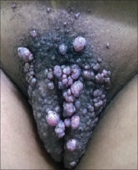
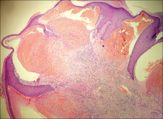
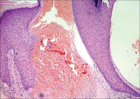
Angiokeratomas are benign tumors characterized by proliferation and dilatation of blood vessels in the upper dermis. They are divided into widespread and localized forms. The localized forms are further classified as angiokeratoma of Fordyce, angiokeratoma circumscriptum neviforme, circumscribed angiokeratoma, and angiokeratoma of Mibelli, of which angiokeratoma of Fordyce is the most common. A 38-year-old female, with no systemic comorbidities presented with recurrent, asymptomatic dark, raised lesions over the vulva for 15 years, progressively increasing in size and number. There were no similar complaints in the family or spouse. On examination, multiple pedunculated hyperpigmented papules and nodular lesions with a verrucous surface were present over the bilateral labia majora and pubic area. Per speculum examination revealed no abnormalities. The oral, conjunctival, and genital mucosae were normal. There were no similar lesions elsewhere over the body. Histopathological examination revealed marked dilatation of papillary dermal vessels forming large, blood-filled cavernous channels, suggestive of angiokeratoma. The lesions were removed using radiofrequency. We present this case due to the rarity of its occurrence and to emphasize the importance of ruling out nonvenereal causes of genital lesions.
Keywords: Angiokeratoma; nodulocystic; vulva.
Conflict of interest statement
There are no conflicts of interest.
Figures
References
-
- Fogagnolo L, Cintra ML, Velho PE. Angiokeratoma of the vulva. An Bras Dermatol. 2011;86:333–5. - PubMed
-
- Imperial R, Helwig EB. Angiokeratoma. A clinicopathological study. Arch Dermatol. 1967;95:166–75. - PubMed
-
- Blair C. Angiokeratoma of vulva. Br J Dermatol. 1970;83:401–11. - PubMed
-
- Laxmisha C, Dubey AK, Thappa DM, Jayanthi S. Angiokeratoma of fordyce. Indian J Sex Transm Dis. 2004;25:42–3.
Publication types
LinkOut - more resources
Full Text Sources
Molecular Biology Databases
