. 2019 Mar;76(6):1185-1199.
doi: 10.1007/s00018-018-03003-2. Epub 2019 Jan 8.

Monitoring activities of receptor tyrosine kinases using a universal adapter in genetically encoded split TEV assays

Affiliations

Monitoring activities of receptor tyrosine kinases using a universal adapter in genetically encoded split TEV assays

Jan P Wintgens et al. Cell Mol Life Sci. 2019 Mar.

Erratum in

Abstract

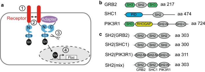
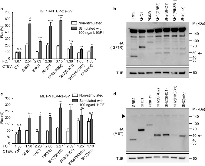
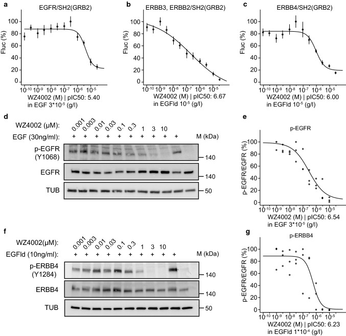
Receptor tyrosine kinases (RTKs) play key roles in various aspects of cell biology, including cell-to-cell communication, proliferation and differentiation, survival, and tissue homeostasis, and have been implicated in various diseases including cancer and neurodevelopmental disorders. Ligand-activated RTKs recruit adapter proteins through a phosphotyrosine (p-Tyr) motif that is present on the RTK and a p-Tyr-binding domain, like the Src homology 2 (SH2) domain found in adapter proteins. Notably, numerous combinations of RTK/adapter combinations exist, making it challenging to compare receptor activities in standardised assays. In cell-based assays, a regulated adapter recruitment can be investigated using genetically encoded protein-protein interaction detection methods, such as the split TEV biosensor assay. Here, we applied the split TEV technique to robustly monitor the dynamic recruitment of both naturally occurring full-length adapters and artificial adapters, which are formed of clustered SH2 domains. The applicability of this approach was tested for RTKs from various subfamilies including the epidermal growth factor (ERBB) family, the insulin receptor (INSR) family, and the hepatocyte growth factor receptor (HGFR) family. Best signal-to-noise ratios of ligand-activated RTK receptor activation was obtained when clustered SH2 domains derived from GRB2 were used as adapters. The sensitivity and robustness of the RTK recruitment assays were validated in dose-dependent inhibition assays using the ERBB family-selective antagonists lapatinib and WZ4002. The RTK split TEV recruitment assays also qualify for high-throughput screening approaches, suggesting that the artificial adapter may be used as universal adapter in cell-based profiling assays within pharmacological intervention studies.

Keywords: Cell-based assay; Lapatinib; Receptor tyrosine kinases; Split TEV recruitment assay; TEV protease.


Conflict of interest statement

The authors declare competing financial interest.

Figures

Fig. 1
Fig. 1
Design of a versatile split TEV recruitment assay for receptor tyrosine kinases. a Scheme of the split TEV recruitment assay for receptor tyrosine kinases (RTKs). RTKs are fused to an NTEV moiety along with a TEV protease cleavage site (tcs) and an artificial co-transcriptional activator GAL4-VP16 (GV). Adapter proteins are fused to CTEV. Upon activation by a specific ligand (1), the RTK dimerises, is cross-phosphorylated by the kinase domains at Tyr residues, providing docking sites for adapter proteins that bind to phosphorylated tyrosines (2). The ligand-induced interaction between RTK and adapter causes the NTEV and CTEV moieties to form a reconstituted TEV protease (2). Reconstituted TEV protease cleaves at tcs to release GV (3). Liberated GV migrates to the nucleus and initiates expression of firefly luciferase (Fluc) (4). b Domain organisation of full-length adapter proteins that are recruited by ERBB receptors. SH2 src homology 2 domain, SH3 src homology 3 domain, PID phosphotyrosine interaction domain, RHOGAP RhoGAP domain. Note that the adapter PIK3R1 contains two SH2 domains denoted as SH2-N (N-terminal) and SH2-C (C-terminal). c Domain organisation of the artificially concatenated SH2 domain phospho-adapters. For each clustered SH2 adapter, three single SH2 domains were fused. The SH2(mix) adapter contains an SH2 domain taken from each full-length adapter depicted in (b)
Fig. 2
Fig. 2
Comparing adapter protein performance for split TEV recruitment assays to monitoring ERBB receptor activities. Split TEV recruitment assays for ERBB family receptors. EGFR (a), ERBB2/ERBB3 (c), and ERBB4 (e) activities were assessed in PC12 cells using EGF to stimulate EGFR, and EGF-like domain (EGFld) to stimulate ERBB3 and ERBB4. For split TEV assays, the indicated receptor fusions were transfected together with indicated adapters that were fused to the CTEV moiety. Note that for the ERBB2/ERBB3 assay (c), ERBB2 is co-transfected to allow heterodimerisation and thus ERBB3 phosphorylation, which is required for the recruitment of adapters. Assays were stimulated for 16 h and analysed by a firefly luciferase assay. Non-stimulated samples are shown as open bars and stimulated ones as grey bars. FC fold change, Ctrl control (no adapter transfected). Results are shown as average of six samples, and error bars are shown as SEM. Significance was calculated using the unpaired t test, with **p ≤ 0.01; ***p ≤ 0.001; ****p ≤ 0.0001; n.s. not significant. Precise p values are provided in Table S1. Biochemical validation of the expression of ERBB receptors and adapters. Plasmids encoding EGFR (b), ERBB3 (d), and ERBB4 (f) (all tagged with NTEV-tcs-GV-2HA), ERBB2-V5 (d), and adapter proteins (all tagged with CTEV-2HA) were transiently transfected into PC12 cells, allowed to express for 16 h, and lysed. Lysates were subjected to Western blotting using the indicated antibodies. Calculated sizes of fusion proteins are provided in Table S1. Arrow indicates bands of artificial adapter fusions. Note that SH2(PIK3R1) is only very weakly expressed
Fig. 3
Fig. 3
Comparing adapter protein performance for split TEV recruitment assays to monitoring IGF1R and MET receptor activities. a, c Split TEV recruitment assays for IGF1R and MET receptors. IGF1R (a) and MET (c) activities were assessed in PC12 cells using IGF1 to stimulate IGF1R, and HGF to stimulate MET. For split TEV assays, the indicated receptor fusions were transfected together with indicated adapters that were fused to the CTEV moiety. Assays were stimulated for 16 h and analysed by a firefly luciferase assay. Non-stimulated samples are shown as open bars and stimulated ones as grey bars. FC fold change, Ctrl control (no adapter transfected). Results are shown as average of six samples, error bars are shown as SEM. Significance was calculated using the unpaired t test, with **p ≤ 0.01; ***p ≤ 0.001; ****p ≤ 0.0001; n.s. not significant. Precise p values are provided in Table S1. b, d Biochemical validation of the expression of IGF1R and MET receptors and adapters. Plasmids encoding IGFR1 (b), and MET (d) (all tagged with NTEV-tcs-GV-2HA) and adapter proteins (all tagged with CTEV-2HA) were transiently transfected into PC12 cells, allowed to express for 16 h, and lysed. Lysates were subjected to Western blotting using the indicated antibodies. Calculated sizes of fusion proteins are provided in Table S1. Arrow indicates bands of artificial adapter fusions. Note that SH2(PIK3R1) is only very weakly expressed. Arrowhead indicates band for MET, which is also very weakly expressed
Fig. 4
Fig. 4
The concatenated SH2(GRB2) domain fusion is a universal adapter to profile ERBB, IGF1R, and MET activities. Split TEV recruitment assays using increasing concentrations of agonists (EGF, EGFld, IGF1 and HGF shown below x axis) for the receptors EGFR (a), ERBB2/ERBB3 (b), ERBB4 (c), IGF1R (d), and MET (e). Each receptor fusion (NTEV-tcs-GV tag for EGFR, ERBB3, ERBB4, IGF1R, MET; V5-tagged ERBB2) plasmid was co-transfected with the SH2(GRB2)-CTEV adapter plasmid into PC12 cells. Error bars are shown as SEM, with six replicates per condition
Fig. 5
Fig. 5
The universal SH2(GRB2) adapter displays the highest sensitivity to ERBB family inhibition by lapatinib. Split TEV recruitment assays monitoring the lapatinib-mediated inhibition of EGFR (a), ERBB2/ERBB3 (b), ERBB4 (c), IGF1R (d), and MET (e). Each receptor fusion (NTEV-tcs-GV tag for EGFR, ERBB3, ERBB4, IGF1R, MET; V5-tagged ERBB2) plasmid is co-transfected with the universal SH2(GRB2)-CTEV adapter plasmid into PC12 cells. Depicted are dose–response curves with a constant agonist stimulus (EGF, EGFld, IGF1, and HGF) and increasing concentrations of lapatinib. Error bars are shown as SEM, with six replicates per condition. f Heatmap displaying pIC50 values for lapatinib comparing assay performance of full-length and SH2(GRB2) domain concatenated CTEV adapters co-transfected with the ERBB family NTEV-tcs-GV fusions. Lapatinib reduces p-EGFR (Y1068) levels in A549 cells (g, h) and p-ERBB4 (Y1284) in T-47D cells (i, j). Cells were treated for 1 h with increasing concentrations of lapatinib and stimulated for 5 min with 30 ng/ml EGF (g) or 10 ng/ml EGFld (i) where indicated. Lysates were subjected to Western blotting and probed with indicated antibodies. Quantification of p-EGFR/EGFR levels (h) as shown in (g) and p-ERBB4/ERBB4 levels (j) as shown in (i) are plotted as dose–response curves. For each concentration depicted, three data points from three different lysates were used for calculations (c.f. Fig. S11a, b)
Fig. 6
Fig. 6
The ERBB family antagonist WZ4002 inhibits split TEV recruitment assays using the universal SH2(GRB2) adapter. Split TEV recruitment assays monitoring the WZ4002-mediated inhibition of EGFR (a), ERBB2/ERBB3 (b), and ERBB4 (c). Each receptor fusion (NTEV-tcs-GV tag for EGFR, ERBB3, ERBB4; V5-tagged ERBB2) plasmid is co-transfected with the universal SH2(GRB2) CTEV adapter into PC12 cells. Depicted are dose–response curves with a constant stimulus (EGF, EGFld) and increasing concentrations of lapatinib. Error bars are shown as SEM, with six replicates per condition. WZ4002 reduces p-EGFR (Y1068) levels in A549 cells (d, e) and p-ERBB4 (Y1284) in T-47D cells (f, g). Cells were treated for 1 h with increasing concentrations of WZ4002 and stimulated for 5 min with 30 ng/ml EGF (d) or 10 ng/ml EGFld (f) where indicated. Lysates were subjected to Western blotting and probed with the indicated antibodies. Quantification of p-EGFR/EGFR levels (e) as shown in (d) and p-ERBB4/ERBB4 levels (g) as shown in (f) are plotted as dose–response curves. For each concentration depicted, three data points from three different lysates were used for calculations (c.f. Fig. S11c, d)

References

    1. Blume-Jensen P, Hunter T. Oncogenic kinase signalling. Nature. 2001;411:355–365. doi: 10.1038/35077225. - DOI - PubMed
    1. Lemmon MA, Schlessinger J. Cell signaling by receptor tyrosine kinases. Cell. 2010;141:1117–1134. doi: 10.1016/j.cell.2010.06.011. - DOI - PMC - PubMed
    1. Mei L, Nave K-A. Neuregulin-ERBB signaling in the nervous system and neuropsychiatric diseases. Neuron. 2014;83:27–49. doi: 10.1016/j.neuron.2014.06.007. - DOI - PMC - PubMed
    1. Santos R, Ursu O, Gaulton A, et al. A comprehensive map of molecular drug targets. Nat Rev Drug Discov. 2017;16:19–34. doi: 10.1038/nrd.2016.230. - DOI - PMC - PubMed
    1. Yaffe MB. Phosphotyrosine-binding domains in signal transduction. Nat Rev Mol Cell Biol. 2002;3:177–186. doi: 10.1038/nrm759. - DOI - PubMed

MeSH terms