

Abnormalities of white matter microstructure in unmedicated patients with obsessive-compulsive disorder: Changes after cognitive behavioral therapy
- PMID: 30623612
- PMCID: PMC6379596
- DOI: 10.1002/brb3.1201
Abnormalities of white matter microstructure in unmedicated patients with obsessive-compulsive disorder: Changes after cognitive behavioral therapy
Abstract
Background: Cognitive behavioral therapy (CBT) is an effective treatment for Obsessive-compulsive disorder (OCD). Structural and functional white matter defects may suggest a vital neurobiological basis of OCD. However, the effects of CBT on white matter in OCD remain unknown.
Objective: The aim was to investigate white matter changes and the effect of CBT on white matter in OCD patients.
Methods: Fractional anisotropy (FA) maps were acquired using DTI. Participants included 85 patients with OCD and 90 healthy controls. VBM was then performed to detect regions with significant group differences.
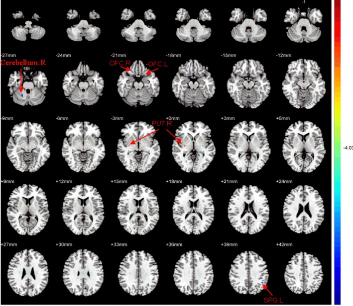
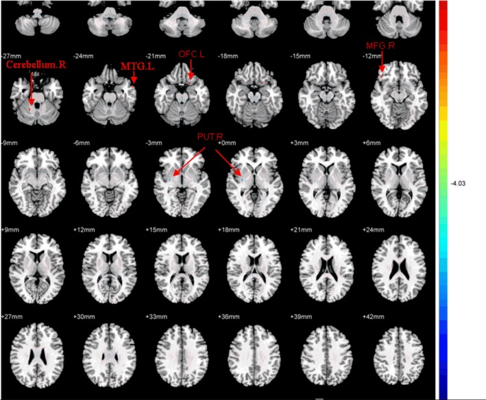
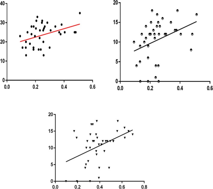
Results: Obsessive-compulsive disorder patients exhibited significantly reduced FA values in bilateral OFC, right cerebellum, and left SPG, while higher FA values were observed in right PUT compared with healthy controls. Following CBT, OCD patients showed higher FA values in right MFG, left OFC, right cerebellum, and left MTG, and decreased FA values in right PUT in comparison with pretreatment. Furthermore, FA values in the left OFC of patients were significantly positively correlated with the Y-BOCS and its associated Compulsions subscale, and FA values in the right PUT were positively correlated with Compulsions subscale. In addition, the percentage change in FA values in left MTG was positively correlated with the percentage reduction in Compulsions subscale, while the percentage change in FA values in left OFC and right PUT was negatively correlated with the percentage reductions in Obsessive and Compulsions subscale, respectively.
Conclusions: Our findings demonstrate the abnormalities of white matter microstructure in unmedicated patients with OCD. These abnormalities may be partly reversed by CBT.
Keywords: cognitive behavioral therapy; diffusion tensor imaging; obsessive-compulsive disorder; white matter.
© 2019 The Authors. Brain and Behavior published by Wiley Periodicals, Inc.
Figures
References
-
- Banca, P. , Voon, V. , Vestergaard, M. D. , Philipiak, G. , Almeida, I. , Pocinho, F. , … Castelo‐Branco, M. (2015). Imbalance in habitual versus goal directed neural systems during symptom provocation in obsessive‐compulsive disorder. Brain, 138(3), 798–811. 10.1093/brain/awu379 - DOI - PMC - PubMed
-
- Benedetti, F. , Giacosa, C. , Radaelli, D. , Poletti, S. , Pozzi, E. , Dallaspezia, S. , … Smeraldi, E. (2013). Widespread changes of white matter microstructure in obsessive‐compulsive disorder: Effect of drug status. European Neuropsychopharmacology, 23(7), 581–593. 10.1016/j.euroneuro.2012.07.002 - DOI - PubMed
MeSH terms
LinkOut - more resources
Full Text Sources
Medical
