Review
. 2019 May 8;119(9):6184-6226.
doi: 10.1021/acs.chemrev.8b00460. Epub 2019 Jan 9.

Computational Modeling of Realistic Cell Membranes

Affiliations
Review

Computational Modeling of Realistic Cell Membranes

Siewert J Marrink et al. Chem Rev. .

Abstract

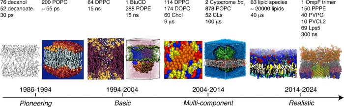
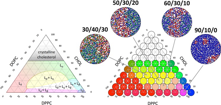
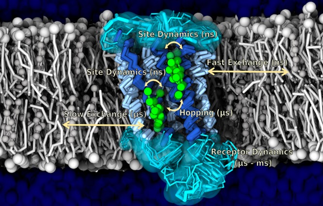
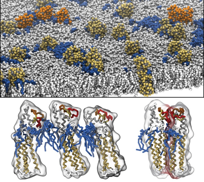
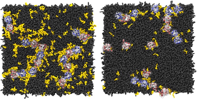
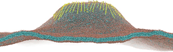
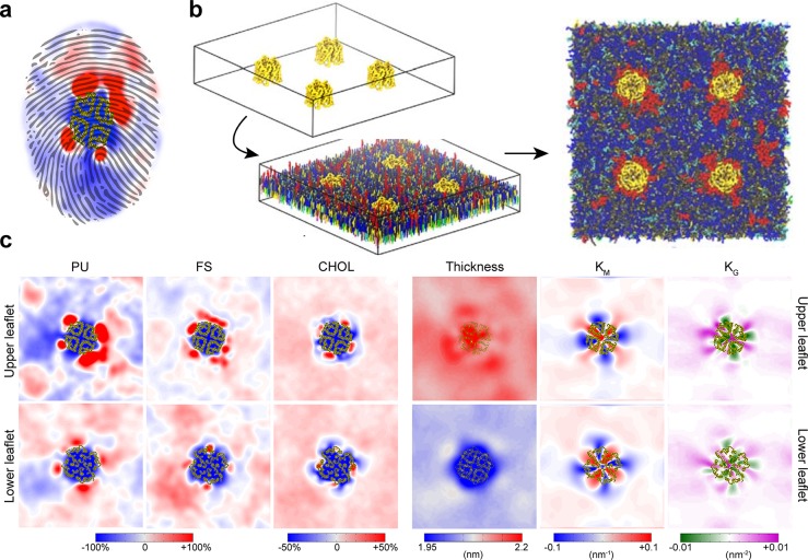
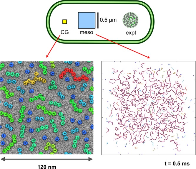
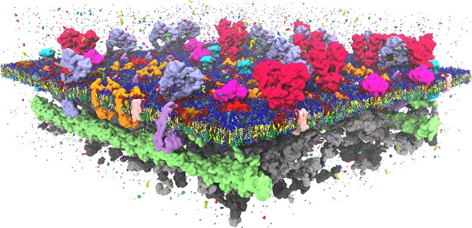
Cell membranes contain a large variety of lipid types and are crowded with proteins, endowing them with the plasticity needed to fulfill their key roles in cell functioning. The compositional complexity of cellular membranes gives rise to a heterogeneous lateral organization, which is still poorly understood. Computational models, in particular molecular dynamics simulations and related techniques, have provided important insight into the organizational principles of cell membranes over the past decades. Now, we are witnessing a transition from simulations of simpler membrane models to multicomponent systems, culminating in realistic models of an increasing variety of cell types and organelles. Here, we review the state of the art in the field of realistic membrane simulations and discuss the current limitations and challenges ahead.


Conflict of interest statement

The authors declare no competing financial interest.

Figures

Figure 1
Figure 1
Growth of complexity of membrane models. From the pioneering stage 30 years ago, basic properties of one and two component membranes were explored around the millennium. From then on, complexity of simulated membrane systems was gradually increased, culminating in the current era of more and more realistic membrane models. POPC, 1-palmitoyl-2-oleoyl-sn-glycero-3-phosphocholine; DPPC, 1,2-dipalmitoyl-sn-glycero-3-phosphocholine; POPE, 1-palmitoyl-2-oleoyl-sn-glycero-3-phosphoethanolamine; DOPC, 1,2-dioleoyl-sn-glycero-3-phosphocholine; Chol, cholesterol; CLs, cardiolipins; PPPE, 1-palmitoyl-2-palmitoleoyl-phosphatidylethanolamine; PVPG, 1-palmitoyl-2-vacenoyl-phosphatidylglycerol; PVCL2, 1,10-palmitoyl-2,20-vacenoyl cardiolipin; Lps5, E. coli R1 lipopolysaccharide core with repeating units of O6-antigen. From left to right: Reprinted with permission from ref (20). Copyright 1988 AIP Publishing. Adapted from ref (26). Copyright 1993 American Chemical Society. Adapted from ref (42). Copyright 2001 American Chemical Society. Adapted with permission from ref (38). Copyright 2004 American Society for Biochemistry and Molecular Biology. Adapted from ref (311). Copyright 2014 American Chemical Society. Adapted from ref (382). Copyright 2013 American Chemical Society. Adapted from ref (593). Copyright 2014 American Chemical Society. Adapted with permission from ref (643). Copyright 2016 Elsevier.
Figure 2
Figure 2
Different resolutions in particle-based simulation models of lipid membranes. At the all-atom (AA) level, all atoms are considered explicitly. Upon coarse-graining, small groups of atoms and associated hydrogens are represented by coarse-grain (CG) beads. Moving down in resolution to the supra-CG level, lipids and proteins are represented only qualitative by few-bead models, and solvent is considered implicitly. Further reduction in resolution is achieved by integrating out also the lipid particles by mean-field approaches.
Figure 3
Figure 3
Example of a multicomponent membrane phase diagram. The ternary lipid mixture dioleoyl phosphocholine (DOPC), DPPC, and cholesterol exhibits a range of interesting phase behavior. Experimental phase diagram is shown on the left, and a simulated diagram using further optimized Martini parameters for DOPC and DPPC on the right. Inserts show snapshots of four of the different simulations illustrating the phase separation. The lipids are colored red, blue, and green for DOPC, DPPC, and cholesterol, respectivly. Adapted from ref (335). Copyright 2018 American Chemical Society.
Figure 4
Figure 4
Example of protein–lipid binding modes. Cholesterol binding to a GPCR with indications of fast and slow exchange dynamics, obtained from MD simulations by Sengupta and co-workers.
Figure 5
Figure 5
Protein-induced lipid flip-flop. (A) Overlay of POPC lipids bound to Opsin at intermediate stages of the flip-flop pathway. (B) CG trajectory of a DOPG lipid during a (partial) flip-flop mediated by the SecYEG complex. (C) Overlay of POPC configurations bound to a fungal scamblase, defining a flip-flop pathway. In all snapshots, the lipid phosphate groups are represented by red spheres. The tails are colored from yellow to green to visualize the flip-flop pathway.
Figure 6
Figure 6
Example of lipid-mediated protein–protein oligomerization. The snapshots show how gangliosides mediate cluster formation of tetraspanin CD81 proteins. Reproduced from ref (497). Copyright 2014 American Chemical Society.
Figure 7
Figure 7
Example of lipid-mediated protein clustering. Snapshots of clustering of GPCRs in a healthy membrane (left), containing DHA, and a nonhealthy membrane (right) depleted of DHA. In the healthy case, higher order oligomers, stabilized by DHA, are more present. Reproduced with permission from ref (501). Copyright 2016 Nature (http://creativecommons.org/licenses/by/4.0/).
Figure 8
Figure 8
Membrane curvature generation by proteins. Onset of membrane tubulation induced by 48 copies of α-Syn100 (yellow) interacting with a membrane composed of 85296 POPG lipids (blue tails, red headgroups). Water is not shown for clarity. Snapshot is obtained at 300 ns simulation time with the Martini model. The budding tubule extends ∼25 nm above the bulk lipid bilayer. Adapted from ref (533). Copyright 2014 American Chemical Society.
Figure 9
Figure 9
An example of a complex plasma membrane model. Corradi et al. simulated ten different membrane proteins in a 63 lipid PM mixture. Each protein’s different TM shape and lipid–protein interactions resulted in a unique lipid fingerprint (a). Here AQP1 is depicted showing the simulation setup, snapshot of the (b) outer membrane as well as lipid enrichment/depletion and bilayer properties around the (c) protein. Adapted from ref (603). Copyright 2018 American Chemical Society.
Figure 10
Figure 10
Example of a complex bacterial membrane model. Showing the outer and inner E. coli cell membrane with embedded membrane proteins including the membrane spanning multidrug efflux pump AcrABZ-TolC. Adapted from ref (653). Copyright 2017 American Chemical Society.
Figure 11
Figure 11
Developing a mesoscale model for simulation of bacterial outer membrane protein islands. The top panel shows a schematic diagram of an E. coli cell, with the areas of outer membrane studied via CG simulation (yellow square), by mesoscale simulation (blue square), and by experimental single molecule tracking (green circle) shown to scale. The lower two panels are snapshots from CG (left) and meso (right) simulations of OMP clustering (see main text and ref (688) for details).
Figure 12
Figure 12
Glimpse at the near future: model of the plasma membrane in full complexity. Featuring: a lipid bilayer composed of hundreds of different lipids, crowded with a large variety of embedded as well as peripherally bound proteins, a supporting actin skeleton, a cytoplasmic site full of proteins, and realistic gradients of metabolites, ions, and pH.

References

    1. Yang Y.; Lee M.; Fairn G. D. Phospholipid Subcellular Localization And Dynamics. J. Biol. Chem. 2018, 293, 6230–6240. 10.1074/jbc.R117.000582. - DOI - PMC - PubMed
    1. Harayama T.; Riezman H. Understanding The Diversity Of Membrane Lipid Composition. Nat. Rev. Mol. Cell Biol. 2018, 19, 281–296. 10.1038/nrm.2017.138. - DOI - PubMed
    1. Van Meer G.; Voelker D. R.; Feigenson G. V. Membrane Lipids: Where They Are And How They Behave. Nat. Rev. Mol. Cell Biol. 2008, 9, 112–24. 10.1038/nrm2330. - DOI - PMC - PubMed
    1. Lingwood D.; Simons K. Lipid Rafts As A Membrane-Organizing Principle. Science 2010, 327, 46–50. 10.1126/science.1174621. - DOI - PubMed
    1. Jacobson K.; Mouritsen O. G.; Anderson R. G. Lipid Rafts: At A Crossroad Between Cell Biology And Physics. Nat. Cell Biol. 2007, 9, 7–14. 10.1038/ncb0107-7. - DOI - PubMed

Publication types