. 2019 Apr 1;316(4):F682-F692.
doi: 10.1152/ajprenal.00222.2018. Epub 2019 Jan 9.

Uropathogenic Escherichia coli-induced fibrosis, leading to lower urinary tract symptoms, is associated with type 2 cytokine signaling

Affiliations

Uropathogenic Escherichia coli-induced fibrosis, leading to lower urinary tract symptoms, is associated with type 2 cytokine signaling

Ashlee Bell-Cohn et al. Am J Physiol Renal Physiol. .

Abstract

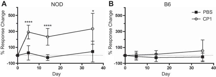
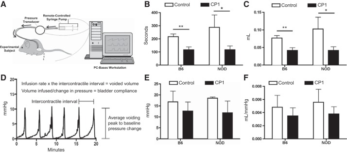
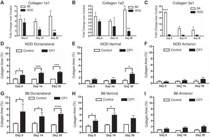
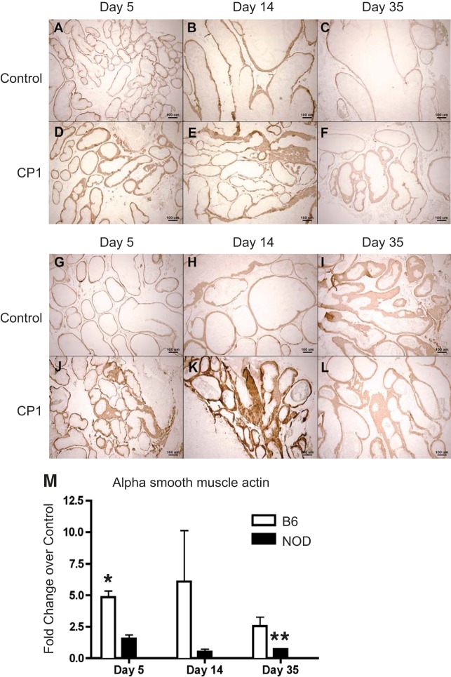
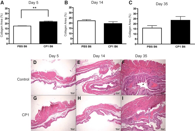
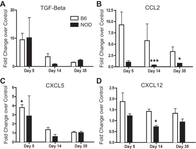
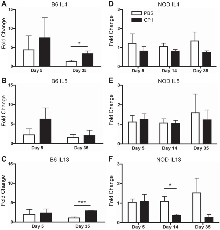
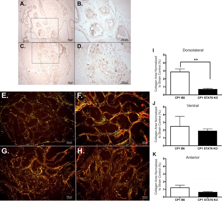
Chronic inflammation and prostate fibrosis have been identified as contributors to lower urinary tract symptoms (LUTS) pathophysiology in humans. It has been shown that transurethral infection of an Escherichia coli strain named CP1, which was isolated from a patient with chronic prostatitis, can lead to the develop of differential chronic inflammation and pain in certain mouse strains. Therefore, we hypothesized that differential inflammation would influence fibrotic response in the prostate. This study showed that while prostatic infection by CP1 causes the development of chronic tactile allodynia in NOD/ShiltJ (NOD) but not C57BL/6 (B6) mice, both mice developed evidence of prostate inflammation, prostate fibrosis, and urinary dysfunction. Fibrosis was confirmed by the upregulation of fibrosis-associated messenger RNAs (mRNAs), α-smooth muscle actin immunohistochemistry, and collagen staining with picrosirius red. These findings were mainly focused on the dorsolateral lobes of the prostate. Both mouse strains also developed smaller, more frequent voiding patterns postinfection, examined via cystometry. B6 mice responded to CP1 infection with type 2 cytokines (IL-4 and IL-13), while NOD mice did not, which may explain the differing tactile allodynia responses and level of collagen deposition. When mice lacking signal transducer and activator of transcription 6 (STAT6), a transcription factor known to be important for the production and signaling of IL-4 and IL-13, were infected with CP1, fibrosis was attenuated. This study provides a potential model for studying the development of infection-induced prostatic fibrosis and LUTS. This study also demonstrates that CP1-induced prostate fibrosis has a STAT6-dependent mechanism in B6 mice.

Keywords: LUTS; UPEC; fibrosis; type 2 cytokine; urinary dysfunction.


Conflict of interest statement

No conflicts of interest, financial or otherwise, are declared by the authors.

Figures

Fig. 1.
Fig. 1.
CP1-infected NOD mice, but not infected B6 mice, have increased tactile allodynia. A: percent response change in control and CP1-infected NOD tactile allodynia responses, normalized to baseline, at days 5, 14, and 35. The dashed line indicates where there is 0 response change. B: percent response change in control and CP1-infected B6 tactile allodynia responses, normalized to baseline, at days 5, 14, and 35. The dashed line indicates where there is 0 response change. The experiment was performed with 4–5 mice per time point per group. Data represent the average total response change of all 5 filaments. Results were analyzed for statistical significance by unpaired t-test. A P < 0.05 was considered statistically significant: ****P < 0.0001.
Fig. 2.
Fig. 2.
NOD and B6 mice develop chronic inflammation after CP1 infection. Day 35 control (A) and CP1 (B) B6 mouse prostates stained with hematoxylin and eosin (H&E). Day 35 control (C) and CP1 (D) NOD mouse prostates stained with H&E. E: quantification of H&E-based inflammation scoring in B6 mice. F: quantification of H&E-based inflammation scoring in NOD mice. The experiment was performed with at least 3 mice per time point per group. Results were analyzed for statistical significance by unpaired t-test. A P < 0.05 was considered statistically significant: **P < 0.01, ***P < 0.001.
Fig. 3.
Fig. 3.
CP1 infection induces urinary dysfunction in mice. A: schematic depiction of the cystometry apparatus. [Reproduced with permission of Med Associates Inc.] B: average amount of time, in seconds, between micturition events in control and CP1-infected B6 and NOD mice at day 35. C: average volume of fluid expelled from control and CP1-infected B6 and NOD mice during micturition at day 35. D: sample cystometric trace that displays how each urinary parameter was calculated. E: average change in pressure during micturition in control and CP1-infected B6 and NOD mice at day 35. F: average voided volume from control and CP1-infected B6 and NOD divided by the average change in pressure, which conveys bladder compliance at day 35. The experiment was performed with 4 mice per group. Results were analyzed for statistical significance by unpaired t-test. A P < 0.05 was considered statistically significant: *P < 0.05, **P < 0.01.
Fig. 4.
Fig. 4.
NOD and B6 mice develop fibrosis after CP1 infection. CP1-infected B6 and NOD prostate mRNA levels of collagen 1a1 (A), collagen 1a2 (B), and collagen 3a1 (C). Control and CP1-infected NOD collagen content in the dorsolateral (D), ventral (E), and anterior (F) prostate lobes at days 5, 14, and 35 based on picrosirius analysis. Control and CP1-infected B6 collagen content in the dorsolateral (G), ventral (H), and anterior (I) prostate lobes at days 5, 14, and 35 based on picrosirius analysis. The experiment was performed with 4 mice per time point per group. Results were analyzed for statistical significance by unpaired t-test. A P < 0.05 was considered statistically significant: *P < 0.05, **P < 0.01, ***P < 0.001, ****P < 0.0001.
Fig. 5.
Fig. 5.
Infected B6 and NOD mice express elevated α-smooth muscle actin (A-SMA) mRNA and protein in the prostate. Immunohistochemical staining of control and CP1-infected B6 (AF) and NOD (GL) prostate protein levels of A-SMA, respectively. CP1-infected B6 and NOD prostate mRNA levels of A-SMA (M) displayed a fold-change over control at days 5, 14, and 35. The experiment was performed with at least 3 mice per time point per group. Results are expressed as means ± SD. Results were analyzed for statistical significance by unpaired t-test. A P < 0.05 was considered statistically significant: *P < 0.05, **P < 0.01.
Fig. 6.
Fig. 6.
CP1-infected B6 mice have elevated bladder collagen levels only at day 5 and similar levels of bladder inflammation. Collagen deposition levels in the bladders of control and CP1-infected B6 mice after 5 days (A), 14 days (B), and 35 days (C) analyzed by picrosirius staining. Days 5, 14, and 35 (DF) control and CP1 (GI) B6 mouse bladders stained with H&E. The experiment was performed with at least 4 mice per group. Results were analyzed for statistical significance by unpaired t-test. A P < 0.05 was considered statistically significant: **P < 0.01.
Fig. 7.
Fig. 7.
Infected B6 and NOD mice express elevated fibrosis-associated mRNA in the prostate. CP1-infected B6 and NOD prostate mRNA levels of TGF-β (A), CCL2 (B), CXCL5 (C), and CXCL12 (D) displayed as fold-change over control at days 5, 14, and 35. The experiment was performed with 3 mice per time point per group. Results are expressed as means ± SD. Results were analyzed for statistical significance by unpaired t-test. A P < 0.05 was considered statistically significant: *P < 0.05, ***P < 0.001.
Fig. 8.
Fig. 8.
CP1-infected B6 mice, but not NOD mice, express elevated type 2 cytokine mRNA in the prostate. Control and CP1-infected B6 prostate IL-4 (A), IL-5 (B), and IL-13 (C) mRNA levels at days 5 and 35. Control and CP1-infected NOD prostate IL-4 (D), IL-5 (E), and IL-13 (F) mRNA levels at days 5, 14, and 35. The experiment was performed with 3–4 mice per time point per group. Results were analyzed for statistical significance by unpaired t-test. A P < 0.05 was considered statistically significant: *P < 0.05, ***P < 0.001.
Fig. 9.
Fig. 9.
Activated STAT6 is elevated and required for fibrosis development in CP1-infected B6 mice. Immunohistochemical staining of phosphorylated STAT6 in day 35 control B6 tissue at ×10 (A) and ×20 (B) magnification and CP1-infected B6 tissue at ×10 (C) and ×20 (D) magnification. The dotted-lined box in the ×10 images indicates the region being viewed in the ×20 images. Picrosirius images of the dorsolateral prostate lobe from PBS B6 (E), CP1 B6 (F), PBS STAT6 KO (G), and CP1 STAT6 KO (H) mice at ×10 magnification (imaged under circular polarized light). The quantification of collagen content in the dorsolateral (I), ventral (J), and anterior (K) lobes of infected B6 and STAT6 KO mice normalized to their respective controls after 28–35 days. The experiment was performed with 3–5 mice per group. Results were analyzed for statistical significance by unpaired t-test. A P < 0.05 was considered statistically significant: **P < 0.01.

Comment in

References

    1. Barber AE, Norton JP, Wiles TJ, Mulvey MA. Strengths and limitations of model systems for the study of urinary tract infections and related pathologies. Microbiol Mol Biol Rev 80: 351–367, 2016. doi: 10.1128/MMBR.00067-15. - DOI - PMC - PubMed
    1. Bauman TM, Nicholson TM, Abler LL, Eliceiri KW, Huang W, Vezina CM, Ricke WA. Characterization of fibrillar collagens and extracellular matrix of glandular benign prostatic hyperplasia nodules. PLoS One 9: e109102, 2014. doi: 10.1371/journal.pone.0109102. - DOI - PMC - PubMed
    1. Cantiello F, Cicione A, Salonia A, Autorino R, Tucci L, Madeo I, Damiano R. Periurethral fibrosis secondary to prostatic inflammation causing lower urinary tract symptoms: a prospective cohort study. Urology 81: 1018–1024, 2013. doi: 10.1016/j.urology.2013.01.053. - DOI - PubMed
    1. Darby I, Skalli O, Gabbiani G. Alpha-smooth muscle actin is transiently expressed by myofibroblasts during experimental wound healing. Lab Invest 63: 21–29, 1990. - PubMed
    1. Darnell SE, Hall TL, Tomlins SA, Cheng X, Ives KA, Roberts WW. Histotripsy of the prostate in a canine model: characterization of post-therapy inflammation and fibrosis. J Endourol 29: 810–815, 2015. doi: 10.1089/end.2014.0585. - DOI - PMC - PubMed

Publication types

MeSH terms