. 2019 Mar 21;30(7):820-827.
doi: 10.1091/mbc.E18-09-0592. Epub 2019 Jan 9.

Effects of altering histone posttranslational modifications on mitotic chromosome structure and mechanics

Affiliations

Effects of altering histone posttranslational modifications on mitotic chromosome structure and mechanics

Ronald Biggs et al. Mol Biol Cell. .

Abstract

During cell division, chromatin is compacted into mitotic chromosomes to aid faithful segregation of the genome between two daughter cells. Posttranslational modifications (PTMs) of histones alter compaction of interphase chromatin, but it remains poorly understood how these modifications affect mitotic chromosome stiffness and structure. Using micropipette-based force measurements and epigenetic drugs, we probed the influence of canonical histone PTMs that dictate interphase euchromatin (acetylation) and heterochromatin (methylation) on mitotic chromosome stiffness. By measuring chromosome doubling force (the force required to double chromosome length), we find that histone methylation, but not acetylation, contributes to mitotic structure and stiffness. We discuss our findings in the context of chromatin gel modeling of the large-scale organization of mitotic chromosomes.


Figures

FIGURE 1:
FIGURE 1:
Experimental setup for chromosome micromanipulation, force measurement, and image quantification. (A) Schematic of the single captured chromosome experimental setup. Single chromosomes were captured from mitotic HeLa cells in a custom-made well (Materials and Methods). Capture was performed after lysing the cell membranes with a PBS–Triton-X solution, where the chromosome was captured from the whole genome chromosome bundle (Supplemental Figure S1). Once captured, the chromosome could be stretched for measurements of the doubling force or sprayed with fluorescent antibodies for immunostaining experiments. (B) An example of an experiment to measure the doubling force of a mitotic chromosome. The force (thin pipette on the left) and pull (larger pipette on the right) pipettes were aligned to be roughly perpendicular to the captured chromosomes. The pull pipette then moved away from the force pipette, stretching the chromosome (dashed line). The stretching of the chromosome would cause the force pipette to deflect (thin, rightward arrow) from its original position (thin, vertical line), which was used to calculate the force on the chromosome for the amount of stretch at that point. Chromosome initial length (thick bar; measured by the distance from the center of the pipettes) and diameter (not shown) measured using a still image in ImageJ.
FIGURE 2:
FIGURE 2:
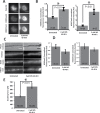
HDACis cause increased H3K9ac fluorescence in mitotic fixed cells and captured chromosomes, but have little effect on the stiffness of mitotic chromosomes. (A) Example representative images of levels of H3K9ac fluorescence measurement on fixed mitotic cells. Scale bar = 10 μm. (B) Quantitative data of A. The H3K9ac intensity ratio of untreated to 2 mM VPA 16–24 h treatment was 1.4 ± 0.1 and is statistically significant. The H3K9ac intensity ratio of untreated to 50 nM TSA 16–24 h treatment was 2.3 ± 0.3 and is statistically significant. (C) Example representative images of levels of H3K9ac fluorescence measurements on captured mitotic chromosomes. Scale bar = 5 μm. (D) Quantitative data of C. The H3K9ac intensity ratio of untreated to 2 mM VPA 16–24 h treatment was 1.8 ± 0.2 and is statistically significant. The H3K9ac intensity ratio of untreated to 50 nM TSA 16–24 h treatment was 2.3 ± 0.6 and is statistically significant. (E) Recorded doubling force for mitotic chromosomes from untreated and HDACi-treated cells. The average chromosome doubling forces were 320 ± 30 pN in untreated cells. The average doubling force was 310 ± 40 pN in 2 mM VPA 16–24 h treated cells, statistically insignificantly different compared with untreated cells. The average doubling force was 330 ± 30 pN in 50 nM TSA 16–24 h treated cells, statistically insignificantly different compared with untreated cells. Error bars represent SE. Asterisk in bar graphs represents a statistically significant difference (p < 0.05). All p values calculated via t test.
FIGURE 3:
FIGURE 3:
Methylstat (HDMi) treatment causes an increase in methylation for mitotic fixed cells and stiffens mitotic chromosomes. (A) Example representative images of levels of H3K9me2,3 and H3K27me3 fluorescence intensity on fixed mitotic cells. Scale bar = 10 μm. (B) Quantitative data of A. The H3K9me2,3 intensity ratio of untreated to 2 μM MS 40–48 h treatment was 1.9 ± 0.1 and is statistically significant. The H3K27me3 intensity ratio of untreated to 2 μM MS 40–48 h treatment was 4.4 ± 0.5 and is statistically significant. (C) Example representative images of levels of H3K9me2,3 and H3K27me3 fluorescence intensity on captured mitotic chromosomes. Scale bar = 5 μm. (D) Quantitative data of C. The H3K9me2,3 intensity ratio of untreated to 2 μM MS 40–48 h treatment was 0.73 ± 0.10, statistically insignificantly different from untreated cells. The H3K27me3 intensity ratio of untreated to 2 μM MS 40–48 h treatment was 0.81 ± 0.09, statistically insignificantly different from untreated cells. (E) Recorded doubling force for mitotic chromosomes from untreated and MS-treated cells. The average chromosome doubling forces were 320 ± 30 pN in untreated cells. The average doubling force was 580 ± 40 pN in 2 μM MS 40–48 h treated cells, a statistically significant increase of ∼80% compared with untreated cells. Error bars represent SE. Asterisk in bar graphs represents a statistically significant difference (p < 0.05). All p values were calculated via t test.
FIGURE 4:
FIGURE 4:
Methylstat treatment does not cause a change in SMC2 fluorescence levels. (A) Example representative images of levels of SMC2 fluorescence intensity on fixed mitotic cells. Scale bar = 10 μm. (B) Quantitative data of A. The SMC2 intensity ratio of untreated to 2 μM MS 40–48 h treatment was 1.1 ± 0.1, statistically insignificantly different from untreated cells. (C) Example representative images of levels of SMC2 fluorescence on captured mitotic chromosomes. Scale bar = 5 μm. (D) Quantitative data of C. The SMC2 intensity ratio of untreated to 2 μM MS 40–48 h treatment was 0.82 ± 0.05, statistically insignificantly different. All p values calculated via t test.
FIGURE 5:
FIGURE 5:
Model of mitotic chromosome. (A) Gel-based model of mitotic chromosomes, demonstrating the cross-linking elements as condensin and the intervening fibers as chromatin. This model is compatible with different models of mitotic chromosomes including the loop-extrusion model, in which condensin can act as both a cross-linking element and the loop-extruding element. (B) Methods in which changes to the chromatin fiber or interactions of the chromatin fiber can stiffen a gel network. These models are not mutually exclusive and can be used to describe how increased histone methylation introduces an increase in stiffness to mitotic chromosomes. Neither of these effects is changed when histones are hyperacetylated in mitosis.

References

    1. Alipour E, Marko JF. (2012). Self-organization of domain structures by DNA-loop-extruding enzymes. Nucleic Acids Res , 11202–11212. - PMC - PubMed
    1. Ball AR, Jr, Yokomori K. (2001). The structural maintenance of chromosomes (SMC) family of proteins in mammals. Chromosome Res , 85–96. - PubMed
    1. Banigan EJ, Stephens AD, Marko JF. (2017). Mechanics and buckling of biopolymeric shells and cell nuclei. Biophys J , 1654–1663. - PMC - PubMed
    1. Beck DB, Oda H, Shen SS, Reinberg D. (2012). PR-Set7 and H4K20me1: at the crossroads of genome integrity, cell cycle, chromosome condensation, and transcription. Genes Dev , 325–337. - PMC - PubMed
    1. Bilokapic S, Strauss M, Halic M. (2018). Cryo-EM of nucleosome core particle interactions in trans. Sci Rep , 7046. - PMC - PubMed

Publication types