

Population-Based Assessment of the Association Between Magnetic Resonance Imaging Background Parenchymal Enhancement and Future Primary Breast Cancer Risk
- PMID: 30625040
- PMCID: PMC6494266
- DOI: 10.1200/JCO.18.00378
Population-Based Assessment of the Association Between Magnetic Resonance Imaging Background Parenchymal Enhancement and Future Primary Breast Cancer Risk
Abstract
Purpose: To evaluate comparative associations of breast magnetic resonance imaging (MRI) background parenchymal enhancement (BPE) and mammographic breast density with subsequent breast cancer risk.
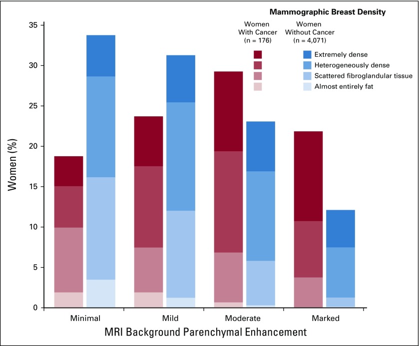
Patients and methods: We examined women undergoing breast MRI in the Breast Cancer Surveillance Consortium from 2005 to 2015 (with one exam in 2000) using qualitative BPE assessments of minimal, mild, moderate, or marked. Breast density was assessed on mammography performed within 5 years of MRI. Among women diagnosed with breast cancer, the first BPE assessment was included if it was more than 3 months before their first diagnosis. Breast cancer risk associated with BPE was estimated using Cox proportional hazards regression.
Results: Among 4,247 women, 176 developed breast cancer (invasive, n = 129; ductal carcinoma in situ,n = 47) over a median follow-up time of 2.8 years. More women with cancer had mild, moderate, or marked BPE than women without cancer (80% v 66%, respectively). Compared with minimal BPE, increasing BPE levels were associated with significantly increased cancer risk (mild: hazard ratio [HR], 1.80; 95% CI, 1.12 to 2.87; moderate: HR, 2.42; 95% CI, 1.51 to 3.86; and marked: HR, 3.41; 95% CI, 2.05 to 5.66). Compared with women with minimal BPE and almost entirely fatty or scattered fibroglandular breast density, women with mild, moderate, or marked BPE demonstrated elevated cancer risk if they had almost entirely fatty or scattered fibroglandular breast density (HR, 2.30; 95% CI, 1.19 to 4.46) or heterogeneous or extremely dense breasts (HR, 2.61; 95% CI, 1.44 to 4.72), with no significant interaction (P = .82). Combined mild, moderate, and marked BPE demonstrated significantly increased risk of invasive cancer (HR, 2.73; 95% CI, 1.66 to 4.49) but not ductal carcinoma in situ (HR, 1.48; 95% CI, 0.72 to 3.05).
Conclusion: BPE is associated with future invasive breast cancer risk independent of breast density. BPE should be considered for risk prediction models for women undergoing breast MRI.
Conflict of interest statement
The statements in this publication are solely the responsibility of the authors and do not necessarily represent the views of the Patient-Centered Outcomes Research Institute or its Board of Governors or Methodology Committee or the views of the National Cancer Institute or the National Institutes of Health.
Figures
Comment in
-
Predict, Then Act: Moving Toward Tailored Prevention.J Clin Oncol. 2019 Apr 20;37(12):943-945. doi: 10.1200/JCO.19.00068. Epub 2019 Mar 7. J Clin Oncol. 2019. PMID: 30844319 No abstract available.
Similar articles
-
Factors Associated With Background Parenchymal Enhancement on Contrast-Enhanced Mammography.AJR Am J Roentgenol. 2021 Feb;216(2):340-348. doi: 10.2214/AJR.19.22353. Epub 2020 Dec 23. AJR Am J Roentgenol. 2021. PMID: 32755162
-
Automated rating of background parenchymal enhancement in MRI of extremely dense breasts without compromising the association with breast cancer in the DENSE trial.Eur J Radiol. 2024 Jun;175:111442. doi: 10.1016/j.ejrad.2024.111442. Epub 2024 Mar 24. Eur J Radiol. 2024. PMID: 38583349
-
Effect of Background Parenchymal Enhancement on Cancer Risk Across Different High-Risk Patient Populations Undergoing Screening Breast MRI.AJR Am J Roentgenol. 2019 Mar 19;212(6):1412-1418. doi: 10.2214/AJR.18.20566. AJR Am J Roentgenol. 2019. PMID: 30888867
-
Impact of Background Parenchymal Enhancement on Diagnostic Performance of Breast MRI: A Systematic Review and Meta-Analysis.Radiology. 2025 May;315(2):e241919. doi: 10.1148/radiol.241919. Radiology. 2025. PMID: 40423535
-
The Association of Background Parenchymal Enhancement at Breast MRI with Breast Cancer: A Systematic Review and Meta-Analysis.Radiology. 2019 Sep;292(3):552-561. doi: 10.1148/radiol.2019182441. Epub 2019 Jun 25. Radiology. 2019. PMID: 31237494
Cited by
-
Association of Breast Cancer Odds with Background Parenchymal Enhancement Quantified Using a Fully Automated Method at MRI: The IMAGINE Study.Radiology. 2023 Sep;308(3):e230367. doi: 10.1148/radiol.230367. Radiology. 2023. PMID: 37750771 Free PMC article.
-
TopoTxR: A topology-guided deep convolutional network for breast parenchyma learning on DCE-MRIs.Med Image Anal. 2025 Jan;99:103373. doi: 10.1016/j.media.2024.103373. Epub 2024 Oct 16. Med Image Anal. 2025. PMID: 39454312 Free PMC article.
-
Applying artificial intelligence technology to assist with breast cancer diagnosis and prognosis prediction.Front Oncol. 2022 Aug 31;12:980793. doi: 10.3389/fonc.2022.980793. eCollection 2022. Front Oncol. 2022. PMID: 36119479 Free PMC article. Review.
-
Clinical Artificial Intelligence Applications: Breast Imaging.Radiol Clin North Am. 2021 Nov;59(6):1027-1043. doi: 10.1016/j.rcl.2021.07.010. Radiol Clin North Am. 2021. PMID: 34689871 Free PMC article. Review.
-
Quantitative evaluation of Kaiser score in diagnosing breast dynamic contrast-enhanced magnetic resonance imaging for patients with high-grade background parenchymal enhancement.Quant Imaging Med Surg. 2023 Oct 1;13(10):6384-6394. doi: 10.21037/qims-23-113. Epub 2023 Aug 17. Quant Imaging Med Surg. 2023. PMID: 37869283 Free PMC article.
