. 2019 Jan 9;15(1):e1007530.
doi: 10.1371/journal.ppat.1007530. eCollection 2019 Jan.

Recruitment of Vps34 PI3K and enrichment of PI3P phosphoinositide in the viral replication compartment is crucial for replication of a positive-strand RNA virus

Affiliations

Recruitment of Vps34 PI3K and enrichment of PI3P phosphoinositide in the viral replication compartment is crucial for replication of a positive-strand RNA virus

Zhike Feng et al. PLoS Pathog. .

Abstract

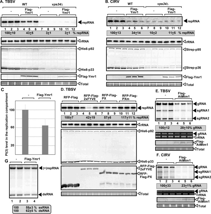
Tombusviruses depend on subversions of multiple host factors and retarget cellular pathways to support viral replication. In this work, we demonstrate that tomato bushy stunt virus (TBSV) and the closely-related carnation Italian ringspot virus (CIRV) recruit the cellular Vps34 phosphatidylinositol 3-kinase (PI3K) into the large viral replication compartment. The kinase function of Vps34 is critical for TBSV replication, suggesting that PI(3)P phosphoinositide is utilized by TBSV for building of the replication compartment. We also observed increased expression of Vps34 and the higher abundance of PI(3)P in the presence of the tombusviral replication proteins, which likely leads to more efficient tombusvirus replication. Accordingly, overexpression of PI(3)P phosphatase in yeast or plants inhibited TBSV replication on the peroxisomal membranes and CIRV replication on the mitochondrial membranes. Moreover, the purified PI(3)P phosphatase reduced TBSV replicase assembly in a cell-free system. Detection of PI(3)P with antibody or a bioprobe revealed the enrichment of PI(3)P in the replication compartment. Vps34 is directly recruited into the replication compartment through interaction with p33 replication protein. Gene deletion analysis in surrogate yeast host unraveled that TBSV replication requires the vesicle transport function of Vps34. In the absence of Vps34, TBSV cannot efficiently recruit the Rab5-positive early endosomes, which provide PE-rich membranes for membrane biogenesis of the TBSV replication compartment. We found that Vps34 and PI(3)P needed for the stability of the p33 replication protein, which is degraded by the 26S proteasome when PI(3)P abundance was decreased by an inhibitor of Vps34. In summary, Vps34 and PI(3)P are needed for providing the optimal microenvironment for the replication of the peroxisomal TBSV and the mitochondrial CIRV.


Conflict of interest statement

The authors have declared that no competing interests exist.

Figures

Fig 1
Fig 1. Vps34p PI3K is an essential host factor for tombusvirus replication in yeast.
(A) Deletion of Vps34 inhibits TBSV replication in yeast. Northern blot analysis of TBSV repRNA using a 3’ end specific probe shows reduced accumulation of repRNA in vps34Δ yeast strain in comparison with the wt yeast strain. Viral proteins His6-p33 and His6-p92 were expressed from plasmids from the galactose-inducible GAL1 promoter, while DI-72(+) repRNA was expressed from the GAL10 promoter. Vps34-Flag and its two defective mutants were separately expressed from a plasmid as shown. Northern blot with 18S ribosomal RNA specific probe was used as a loading control. Bottom images: Western blot analysis of the level of His6-p33 and His6-p92 with anti-His antibody and Vps34p with anti-Flag antibody. (B) Reduced CIRV replication in vps34Δ yeast strain. CIRV proteins Strep-p36 and Strep-p95 were expressed from plasmids from the GAL1 promoter. See further details in panel A.
Fig 2
Fig 2. Chemical inhibitor of Vps34 inhibits TBSV replication and decreases the stability of the viral replication protein.
(A) Inhibition of TBSV replication by the PI3K inhibitor AS604850 in yeast. See further details in Fig 1A. (B) Western blot analysis of TBSV p33 replication protein level in the presence or absence of AS604850 inhibitor. Yeast spheroplasts expressing p33 were treated with MG132 proteasome inhibitor or DMSO as shown. As a loading control, the cellular PGK1 was detected with anti-PGK1 antibody (bottom panel). The samples were taken after 2 h of treatments of spheroplasts. (C) Reduced TBSV RNA production by the tombusvirus replicase assembled in vitro in cell-free extract (CFE) prepared from vps34Δ yeast in comparison with a similar CFE preparation obtained from wt yeast. Purified recombinant p33 and p92pol replication proteins of TBSV and in vitro transcribed TBSV DI-72 (+)repRNA were added to the CFEs. Nondenaturing PAGE analysis shows the 32P-labeled TBSV repRNA products, including the (+)repRNA progeny and the dsRNA replication intermediate, made by the reconstituted TBSV replicase. (D) Reduced TBSV RNA production by the tombusvirus replicase assembled in vitro in CFEs prepared from Wortmannin or DMSO-treated WT yeast. See further details in panel C above. Each experiment was repeated.
Fig 3
Fig 3. Knockdown of VPS34 gene expression inhibits tombusvirus replication in N. benthamiana plants.
(A) Top panel: Accumulation of the TBSV genomic (g)RNA in VPS34- silenced N. benthamiana plants 2 days post-inoculation (dpi) was measured by Northern blot analysis. Inoculation of TBSV gRNA was done 12 days after silencing of VPS34 expression. VIGS was performed via agroinfiltration of tobacco rattle virus (TRV) vector carrying NbVps34 or 3’-terminal GFP (as a control) sequences. Second panel: Ribosomal RNA is shown as a loading control in an ethidium-bromide stained agarose gel. Note that the TBSV genomic RNA is also visible in the gel. Third panel: Northern blot with N. benthamiana 18S ribosomal RNA specific probe was used as a loading control. Fourth panel: RT-PCR analysis of NbVps34 mRNA level in the silenced and control plants. Fifth panel: RT-PCR analysis of TUBULIN mRNA level in the silenced and control plants. Each experiment was repeated. (B) Delayed development of TBSV-induced symptoms is observed in VPS34-silenced N. benthamiana plants as compared with the control plants. Note the lack of phenotype in VPS34-silenced N. benthamiana plants. The picture was taken 5 dpi. (C) Top panel: Accumulation of the CIRV genomic (g)RNA in VPS34-silenced N. benthamiana plants 2.5 dpi was measured by Northern blot analysis. See further details in panel A. (D) Delayed development of CIRV-induced symptoms is observed in VPS34-silenced N. benthamiana plants as compared with the control plants. The picture was taken 5 dpi. (E) Western blot analysis of Vps34 level in yeast replicating TBSV repRNA or lacking viral components. The HA-tagged Vps34p was expressed from its natural promoter and original chromosomal location. The 6xHis-tagged p33 was detected by anti-His antibody. (F) Top panel: Induction of VPS34 mRNA expression in N. benthamiana plants infected with TBSV was detected by semi-quantitative RT-PCR. Middle panel: RT-PCR of tubulin mRNA was used as a control. Bottom panel: Ribosomal RNA is shown as a loading control in an ethidium-bromide-stained agarose gel.
Fig 4
Fig 4. Co-localization of tombusvirus replication protein with Vps34p in yeast.
(A) Confocal laser microscopy images show the partial co-localization of TBSV GFP-tagged p33 replication protein with the RFP-tagged Vps34p protein in wt yeast cells. DIC (differential interference contrast) images are shown on the right. Scale bar represents 2 μm. (B) Super-resolution microscopy images show the partial co-localization of TBSV p33 replication protein with Vps34-FLAG in yeast. Note that p33 was detected with anti-p33 antibody, whereas Vps34 was detected via anti-Flag antibody. Arrows point at the areas where co-localization is observed. Scale bars represent 1 μm. (C) Vps34p is recruited to the large replication compartments consisting of aggregated peroxisomes. The peroxisomes are marked with Pex13-GFP. Scale bars represent 5 μm. (D) Lack of co-localization of Vps34 and Pex13 in the absence of viral replication. See further details in panel C. Scale bars represent 5 μm. (E) Confocal laser microscopy images show the partial co-localization of CIRV GFP-p36 replication protein with Vps34-RFP in yeast. Scale bars represent 5 μm. See further details in panel A.
Fig 5
Fig 5. Recruitment of Vps34 by the TBSV p33 replication protein into the viral replication compartment in N. benthamiana.
(A-B) Confocal microscopy images show co-localization of TBSV p33-BFP replication protein and the AtVps34-GFP, the ortholog of the yeast Vps34 protein, in planta. The large replication compartment was visualized via expression of RFP-SKL peroxisomal matrix marker protein. Expression of the above proteins from the 35S promoter was done after co-agroinfiltration into N. benthamiana leaves. Scale bars represent 5 μm. Note that the top panel in panel B is enlargement of the replication compartment portion of the bottom images. (C) Top images: interaction between TBSV p33-cYFP replication protein and the nYFP-AtVps34 protein was detected by BiFC. Co-localization of RFP-SKL with the BiFC signal (see merged image) demonstrates that the interaction between p33 replication protein and Vps34 occurs in the large viral replication compartments in planta. Control BiFC experiments included p33-cYFP protein in combination with nYFP-MBP protein expressed in N. benthamiana infected with TBSV (bottom images). Scale bars represent 10 μm. (D) Top images: interaction between CIRV p36-cYFP replication protein and the nYFP-AtVps34 protein was detected by BiFC. The CIRV replication compartment was visualized with CoxIV-RFP mitochondria marker. Scale bars represent 10 μm. See further details in panel C.
Fig 6
Fig 6. Interaction between p33 replication protein and Vps34p.
(A) Co-purification of p33 replication protein with the AtVps34-Flag from subcellular membranes of N. benthamiana infected with TBSV. Top two panels: Western blot analysis of co-purified p33 (lane 2) with Flag-affinity purified AtVps34-Flag. P33 was detected with anti-p33 antibody, while AtVps34-Flag was detected with anti-FLAG antibody as shown. Bottom two panels: Western blot of total p33 and AtVps34-Flag in the total plant extracts. (B) Co-purification of p33 and p92pol replication proteins with the yeast Vps34-Flag from subcellular membranes. Top two panels: Western blot analysis of co-purified His6-tagged p33 and His6-p92pol (lanes 2 and 4) with Flag-affinity purified Vps34-Flag. His6-p33 and His6-p92pol were detected with anti-His antibody, while Vps34-Flag was detected with anti-FLAG antibody. The negative control was from yeast expressing Flag peptide and His6-p33 and His6-p92pol purified in a FLAG-affinity column (lanes 1 and 3). Samples were cross-linked or untreated as shown. Middle two panels: Western blot of total His6-p33 and His6-p92pol and Vps34-Flag in the total yeast extracts. Bottom panel: SDS-PAGE analysis of total yeast extract using Coomassie blue staining. (C) Western blot analysis of co-purified 3xHA-tagged Vps34 (lanes 2) with Flag-affinity purified Flag-p33 and Flag-p92pol. Vps34-3xHA was expressed in yeast from its native promoter and original chromosomal location. Vps34-3xHA was detected with anti-HA antibody. The samples were not cross-linked. See further details in panel B above. (D) Pull-down assay including the 2xFlag-tagged yeast Vps34 and its inactive mutants and the MBP-tagged TBSV p33 replication protein. Top panel: Western blot analysis of the captured Vps34p expressed in yeast with the MBP-affinity purified p33 (purified from E. coli) was performed with anti-Flag antibody. The negative control was MBP (lanes 2, 4 and 6). Middle panel: Western blot analysis of the captured MBP-p33 and MBP was performed with anti-MBP antibody. Bottom panel: Western blot analysis of the total 2xFlag-Vps34p in total extracts. (E) Pull-down assay including the GST-tagged yeast Vps34 and its inactive mutant and the MBP-tagged TBSV p33 replication protein. Top panel: Western blot analysis of the captured GST-Vps34p expressed in E. coli with the MBP-affinity purified p33 (from E. coli) was performed with anti-GST antibody. See further details in panel D above. Note that the recombinant GST-Vps34 was purified here, whereas 2xFLAG-Vps34 was present in the soluble fraction of yeast CFE in panel D. (F) Co-purification of Vps34p with the viral replicase shows temporal association. The presence of Vps34p in the membrane-bound viral replicase was tested after blocking cellular translation by cycloheximide. The yeast samples were collected at the shown time points after the addition of cycloheximide to the yeast culture. Note that samples were from yeasts replicating TBSV repRNA. Top panel: Western blot analysis of co-purified 3xHA-tagged Vps34p with Flag-affinity purified Flag-p33 and Flag-p92pol from membrane fraction of yeast. Vps34p was detected with anti-HA antibody. The negative control was His6-tagged p33 and His6-p92pol purified from yeast extracts using a Flag-affinity column. Middle panel: Western blot of purified Flag-p33 detected with anti-Flag antibody. Bottom panels: Western blots of 3xHA-tagged Vps34p, His6-p33 (lane 1) and Flag-p33 proteins in the total yeast extracts using anti-HA, anti-His and anti-Flag antibodies. Each experiment was repeated three times.
Fig 7
Fig 7. Enrichment of PI(3)P into the tombusvirus replication compartment in yeast and in N. benthamiana.
(A) Confocal laser microscopy shows partial co-localization of GFP-tagged p33 replication protein with PI(3)P detected via anti-PI(3)P antibody in yeast cells replicating TBSV repRNA. Peroxisomes were detected with Pex13-BFP marker protein. The bottom panel shows confocal microscopy images of yeast lacking viral components. Scale bars represent 5 μm. (B) Confocal laser microscopy shows partial co-localization of GFP-tagged p33 replication protein with PI(3)P detected via RFP-2xFYVE protein in yeast cells replicating TBSV repRNA. See further details in panel A. Scale bars represent 2 μm. (C) Higher abundance of PI(3)P in yeast replicating TBSV repRNA. PI(3)P abundance was measured with anti-PI(3)P antibody in yeast cells with confocal microscopy and Image J software based on at least 40 cells. Standard deviation is shown. (D) Confocal laser microscopy shows partial co-localization of TBSV BFP-tagged p33 replication protein with PI(3)P detected via anti-PI(3)P antibody in N. benthamiana protoplasts infected with TBSV. The peroxisomes are marked by GFP-SKL. Expression of the above proteins from the 35S promoter was achieved after agroinfiltration into N. benthamiana leaves. Scale bars represent 10 μm. (E) Partial co-localization of p33-BFP, PI(3)P and GFP-AtRab5B protein in N. benthamiana cells infected with TBSV. PI(3)P was detected via expression of RFP-2xFYVE protein. The bottom image shows the localization of PI(3)P and GFP-AtRab5B protein in the mock-infected plant leaves. Scale bars represent 10 μm.
Fig 8
Fig 8. Reduction of PI(3)P inhibits tombusvirus replication in yeasts and plants.
(A) Expression of yeast Ymr1p PI(3)P phosphatase, which produces PI from PI(3)P, inhibits TBSV replication in yeast. Top panel: Northern blot analysis of TBSV repRNA using a 3’ end specific probe shows reduced accumulation of repRNA in WT yeast strain expressing Ymr1p. Viral proteins His6-p33 and His6-p92pol were expressed from plasmids from the GAL1 promoter, while DI-72(+) repRNA was expressed from the GAL10 promoter. Flag-Ymr1p was expressed from a plasmid in WT and vps34Δ yeast strains as shown. Middle panel: Northern blot with 18S ribosomal RNA specific probe was used as a loading control. Bottom images: Western blot analysis of the level of His6-p33 and His6-p92pol with anti-His antibody and Flag-Ymr1p with anti-Flag antibody. (B) Reduced CIRV replication in WT yeast strain expressing of yeast Ymr1p PI(3)P phosphatase. The Strep-tagged CIRV replication proteins were detected with anti-Strep antibody. See further details in panel A. (C) Decreased abundance of PI(3)P in yeast replicating TBSV repRNA and expressing Flag-tagged Ymr1p PI(3)P phosphatase. PI(3)P abundance was measured with anti-PI(3)P antibody within the TBSV replication compartment visualized by expression of GFP-p33 in yeast cells with confocal microscopy and Image J software based on at least 40 cells. Standard deviation is shown. (D) Expression of PI(3)P-binding proteins, which sequester PI(3)P, inhibits TBSV replication in yeast. The WT PX domain and its mutant version (PXm with reduced PI(3)P binding) and the 2xFYVE domain proteins were expressed from high-copy-number plasmids. See further details in panel A. (E) Expression of Flag-tagged AtMtm1 PI(3)P phosphatase, which produces PI from PI(3)P, inhibits TBSV RNA replication in N. benthamiana plants. Top panel: Accumulation of the TBSV genomic (g)RNA in N. benthamiana plants 2 days post-inoculation was measured by Northern blot analysis. Inoculation of TBSV gRNA was done 44 h after agroinfiltration of a plasmid carrying AtMtm1 sequence. Middle panel: ribosomal RNA was used as a loading control. Bottom images: Western blot analysis of the level of Flag-AtMtm1 with anti-Flag antibody. (F) Expression of Flag-tagged AtMtm1 PI(3)P phosphatase inhibits CIRV RNA replication in N. benthamiana plants. See further details in panel E. (G) The affinity purified yeast Ymr1p PI(3)P phosphatase reduces TBSV RNA production by the tombusvirus replicase assembled in vitro in cell-free extract (CFE) prepared from wt yeast. Purified recombinant p33 and p92pol replication proteins of TBSV and in vitro transcribed TBSV DI-72 (+)repRNA were added to the CFEs. Nondenaturing PAGE analysis shows the 32P-labeled TBSV repRNA products, including the (+)repRNA progeny and the dsRNA replication intermediate, made by the reconstituted TBSV replicase. Each experiment was repeated three times.
Fig 9
Fig 9. Components of the Vps34p PI3K complex are essential host factors for tombusvirus replication in yeast.
(A) Deletion of selected yeast genes, which are parts of Vps34 complexes, inhibits TBSV replication in yeast. Top panel: Northern blot analysis of TBSV repRNA using a 3’ end specific probe shows the reduced accumulation of repRNA in the shown yeast strains in comparison with the wt yeast strain. Viral proteins His6-p33 and His6-p92pol were expressed from plasmids from the GAL1 promoter, while DI-72(+) repRNA was expressed from the GAL10 promoter. Middle panel: Northern blot with 18S ribosomal RNA specific probe was used as a loading control. Bottom images: Western blot analysis of the level of His6-p33 and His6-p92pol with anti-His antibody. (B) Deletion of selected yeast genes, which are parts of Vps34 complexes, inhibits CIRV replication in yeast. See further details in panel A. Each experiment was repeated three times.
Fig 10
Fig 10. Vps34p PI3K is required for enrichment of PE within the tombusvirus replication compartment in yeast and in plants.
(A) Lack of recruitment of PI(3)P into the viral replication compartment in the absence of Rab5 GTPase. PI(3)P was detected via expression of RFP-2xFYVE protein in vps21Δypt52Δypt53Δ yeast. (B) Top panels: Confocal laser microscopy images show the lack of enrichment of PE and its lack of co-localization with the TBSV p33/p92 replication proteins in vps34Δ yeast. PE distribution was detected by using biotinylated duramycin peptide and streptavidin conjugated with Alexa Fluor 405. DIC (differential interference contrast) images are shown on the right. Bottom panel: Enrichment of PE and its co-localization with the TBSV p33/p92 replication proteins in wt yeast is visualized by confocal microscopy. Note that yeasts also expressed p92 replication protein and the repRNA. (C) Top panels: VIGS-based knockdown of Vps34 level inhibits the enrichment of PE within the tombusvirus replication compartment in planta. Inoculation of TBSV gRNA was done 12 days after silencing of VPS34 expression. VIGS was performed via agroinfiltration of tobacco rattle virus (TRV) vector carrying NbVps34 sequences. The nonspecific VIGS control was a TRV vector carrying 3’ sequences of GFP (cGFP). Protoplasts were obtained from N. benthamiana leaves, followed by detection of PE distribution with biotinylated duramycin peptide and streptavidin conjugated with Alexa Fluor 405 and confocal microscopy. Bottom images: The experiments were done as in the top panels, except TRV-cGFP was used for VIGS as a control. Each experiment was repeated three times. Scale bars represent 10, 10, 10 and 5 μm (from the top to the bottom panels).
Fig 11
Fig 11. Vps34p PI3K is an essential host factor for several viruses in the Tombusviridae family.
(A-B) Inhibition of TBSV replication by the PI3K inhibitor AS604850 or Wortmannin in N. benthamiana protoplasts. Northern blot analysis of TBSV gRNA using a 3’ end specific probe shows reduced accumulation of gRNA in the inhibitor-treated versus untreated protoplasts. Ethidium-bromide stained agarose gels show ribosomal RNA level as a loading control. (C-D) Northern blot analysis of CIRV gRNA using a 3’ end specific probe shows reduced accumulation of gRNA in the AS604850 or Wortmannin-treated versus untreated protoplasts. See further details in panel A. (E-F) Northern blot analysis of CLSV gRNA using a 3’ end specific probe shows the reduced accumulation of gRNA in the AS604850 or Wortmannin-treated versus untreated protoplasts. See further details in panel A. (G-H) Northern blot analysis of turnip crinkle virus (TCV) gRNA using a 3’ end specific probe shows reduced accumulation of gRNA in the AS604850 or Wortmannin-treated versus untreated protoplasts. See further details in panel A. (I-J) Northern blot analysis of RCNMV RNA1 using a 3’ end specific probe shows reduced accumulation of RNA1 in the AS604850 or Wortmannin-treated versus untreated protoplasts. See further details in panel A.

Similar articles

Cited by

References

    1. Wang A (2015) Dissecting the Molecular Network of Virus-Plant Interactions: The Complex Roles of Host Factors. Annu Rev Phytopathol 53: 45–66. 10.1146/annurev-phyto-080614-120001 - DOI - PubMed
    1. Romero-Brey I, Bartenschlager R (2014) Membranous replication factories induced by plus-strand RNA viruses. Viruses 6: 2826–2857. 10.3390/v6072826 - DOI - PMC - PubMed
    1. Nagy PD (2016) Tombusvirus-Host Interactions: Co-Opted Evolutionarily Conserved Host Factors Take Center Court. Annu Rev Virol 3: 491–515. 10.1146/annurev-virology-110615-042312 - DOI - PubMed
    1. Jordan TX, Randall G (2016) Flavivirus modulation of cellular metabolism. Curr Opin Virol 19: 7–10. 10.1016/j.coviro.2016.05.007 - DOI - PMC - PubMed
    1. Shulla A, Randall G (2016) (+) RNA virus replication compartments: a safe home for (most) viral replication. Curr Opin Microbiol 32: 82–88. 10.1016/j.mib.2016.05.003 - DOI - PMC - PubMed

Publication types

MeSH terms

Substances

Supplementary concepts