. 2019 Jan 8;26(2):330-337.e4.
doi: 10.1016/j.celrep.2018.12.051.

Choline Is an Intracellular Messenger Linking Extracellular Stimuli to IP3-Evoked Ca2+ Signals through Sigma-1 Receptors

Affiliations

Choline Is an Intracellular Messenger Linking Extracellular Stimuli to IP3-Evoked Ca2+ Signals through Sigma-1 Receptors

Eugen Brailoiu et al. Cell Rep. .

Abstract

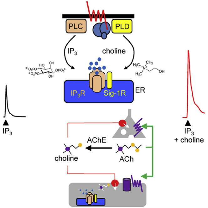
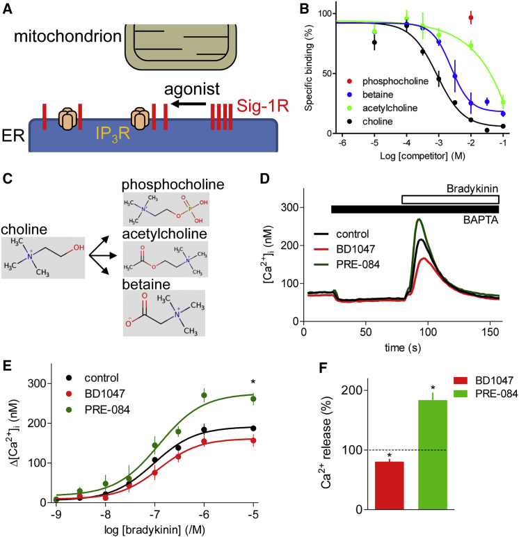
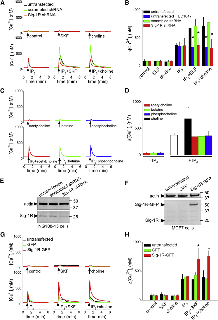
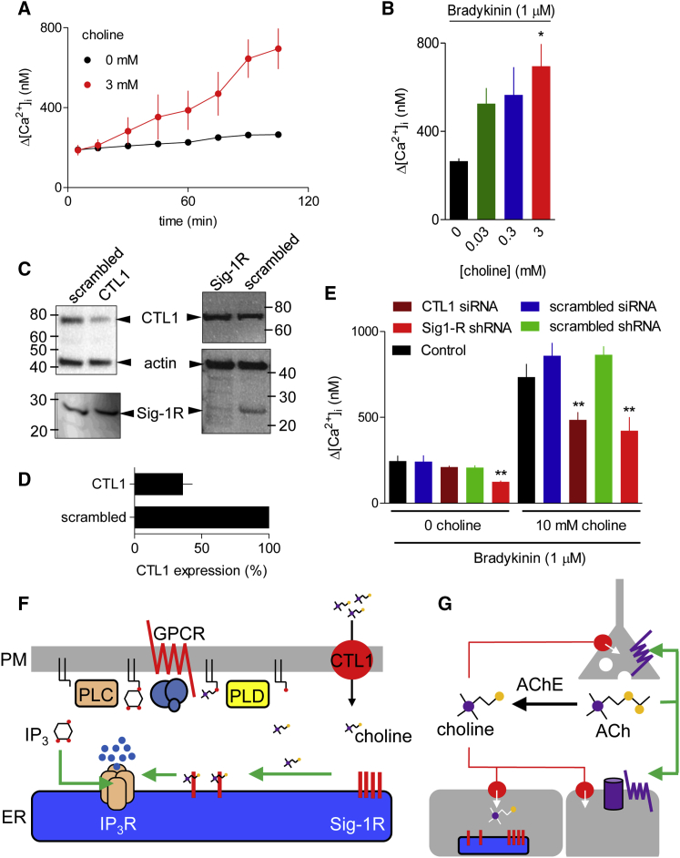
Sigma-1 receptors (Sig-1Rs) are integral ER membrane proteins. They bind diverse ligands, including psychoactive drugs, and regulate many signaling proteins, including the inositol 1,4,5-trisphosphate receptors (IP3Rs) that release Ca2+ from the ER. The endogenous ligands of Sig-1Rs are unknown. Phospholipase D (PLD) cleaves phosphatidylcholine to choline and phosphatidic acid (PA), with PA assumed to mediate all downstream signaling. We show that choline is also an intracellular messenger. Choline binds to Sig-1Rs, it mimics other Sig-1R agonists by potentiating Ca2+ signals evoked by IP3Rs, and it is deactivated by metabolism. Receptors, by stimulating PLC and PLD, deliver two signals to IP3Rs: IP3 activates IP3Rs, and choline potentiates their activity through Sig-1Rs. Choline is also produced at synapses by degradation of acetylcholine. Choline uptake by transporters activates Sig-1Rs and potentiates Ca2+ signals. We conclude that choline is an endogenous agonist of Sig-1Rs linking extracellular stimuli, and perhaps synaptic activity, to Ca2+ signals.

Keywords: Ca(2+); G-protein-coupled receptor; IP(3) receptor; Sigma-1 receptor; bradykinin; choline; intracellular messenger; neurotransmitter; phospholipase C; phospholipase D.


Figures

None
Graphical abstract
Figure 1
Figure 1
Choline Is an Agonist of Sig-1Rs (A) Clusters of Sig-1Rs anchored at MAMs are thought to dissociate into monomers when they bind a Sig-1R agonist, freeing Sig-1Rs to interact with their targets, within and beyond MAMs. The targets include IP3Rs. (B) Specific binding of [3H](+)-pentazocine (5 nM) in the presence of choline and related compounds using membranes from Neuro-2A cells stably expressing Sig-1R-GFP (mean ± SEM; n = 5, with 3 replicates for each). Specific binding of 3H-pentazocine was 90% ± 3% of total binding (mean ± SEM; n = 3) for membranes from cells overexpressing Sig-1R, and 13% ± 5% for mock-transfected cells. (C) Choline metabolism (structures from http://www.hmdb.ca). (D) NG108-15 cells were incubated (2 hr, 37°C) with PRE-084 (25 μM) or BD1047 (25 μM), and then, in the continuous presence of the Sig-1R ligands, loaded with Fluo-8 by incubation with Fluo-8 AM in HEPES-buffered saline (HBS) (30 min, 20°C, with a further 30 min to allow de-esterification of Fluo-8). BAPTA (2.5 mM) was then added to chelate extracellular Ca2+ before addition of bradykinin (10 μM). Results show typical responses as means of 3 replicates. (E) Summary results (mean ± SEM; n = 5, each with 3 replicates) show peak increases in [Ca2+]i (Δ[Ca2+]i) evoked by bradykinin. p < 0.05 for maximal responses relative to control, one-way ANOVA with Dunnett’s test. (F) Pooled results (mean ± SEM; n = 20; as percentages of matched control response) for all bradykinin concentrations. The asterisk () denotes 95% confidence intervals that exclude 100%. See also Figure S1A.
Figure 2
Figure 2
Choline Potentiates IP3-Evoked Ca2+ Release by Stimulating Sig-1Rs (A) Ca2+ signals recorded from Fura-2-loaded NG108-15 cells after microinjection (∼1% cell volume) of IP3 (pipette concentration, 0.5 μM), (+)SKF-10047 (SKF, 100 μM), or choline (100 mM). Results (n = 6 cells) show untransfected cells or after transfection with scrambled shRNA or Sig-1R shRNA, each tagged with red fluorescent protein (RFP). (B) Summary (mean ± SD; n = 6) shows peak [Ca2+]i. p < 0.05, ANOVA with Bonferroni test, relative to matched stimuli in untransfected cells. The effects of pre-incubating cells with BD1047 (25 μM, 15 min) are also shown. (C) Similar analysis of the effects of microinjected IP3 (pipette concentration, 0.5 μM) or acetylcholine, betaine, or phosphocholine (pipette concentration, 100 mM for each), alone or in combination. (D) Summary (mean ± SD; n = 6) shows peak [Ca2+]i. p < 0.05, ANOVA with Bonferroni test, relative to IP3 alone. (E) Western blot (WB) of Sig-1R after transfection of NG108-15 cells with scrambled or Sig-1R shRNA, each tagged with RFP. Tagged shRNAs were used to allow identification of transfected cells in microinjection experiments. Hence, WB from cell populations probably over-estimates Sig-1R expression in functional analyses of micro-injected cells treated with Sig-1R shRNA. Sig-1R expression was reduced to 50% ± 12% of control levels by the shRNA treatment (mean ± SD; n = 3). (F) WB showing detectable expression of Sig-1R in MCF7 cells only after transfection with Sig-1R-GFP. Typical of 4 blots. Mr markers (kDa) are shown. (G) Ca2+ signals recorded from Fura-2-loaded MCF7 cells after microinjection as described for (C). Results (n = 6 cells) are from control cells or after transfection with GFP or Sig-1R-GFP. (H) Summary (mean ± SD; n = 6) results show [ΔCa2+]i. p < 0.05 for maximal responses relative to matched untransfected cells, one-way ANOVA with Dunnett’s test. See also Figures S1B and S1C.
Figure 3
Figure 3
Sig-1R and PLD Contribute to Ca2+ Signals Evoked by Agonists of GPCRs (A) Typical pseudocolor images show peak Ca2+ signals (F340/F380) evoked by ATP (50 μM) in Fura-2-loaded NG108-15 cells transfected with control shRNA or shRNA to PLD1 and PLD2, or Sig-1R, each tagged with RFP. Calibration code (F340/F380) and scale bar (20 μm) apply to all panels. (B) Time course of response to ATP (bar; n = 6). (C) Summary (mean ± SD; n = 6) shows Δ[Ca2+]i evoked by ATP. p < 0.05, ANOVA with Bonferroni test, relative to untransfected cells. (D) Δ[Ca2+]i evoked by bradykinin in populations of NG108-15 cells. Histogram (which shares the y axis) compares responses to bradykinin (10 μM) after treatment with scrambled or Sig-1R shRNA. Results are means ± SEM; n = 3 with duplicate determinations. p < 0.05, Student’s t test. (E) WB shows effects of indicated shRNA, each tagged with RFP, on expression of PLD1 and PLD2 in NG108-15 cells. Mr markers (kDa) are shown. Results, typical of 3 WBs, underestimate knockdowns in the cells used for Ca2+ measurements, which used only cells shown to be transfected by expression of RFP (see A). (F and G) Intracellular concentrations of choline (F) and IP3 (G) during stimulation of NG108-15 cells with ATP (50 μM, bar) show the effects of shRNA for PLD1 and PLD2. Results show means ± SD; n = 6. (H) GPCRs that activate PLC and phospholipase D (PLD) initiate two parallel signaling pathways that converge at IP3Rs. IP3 from PLC directly activates IP3R. Choline from PLD activates Sig-1R, which potentiates IP3-evoked Ca2+ release.
Figure 4
Figure 4
CTL1-Mediated Choline Uptake Potentiates IP3-Evoked Ca2+ Signals (A) NG108-15 cells were incubated in HBS alone or with 3 mM choline for the indicated times before adding bradykinin (1 μM) and immediately recording the increase in [Ca2+]i. Results (mean ± SEM; n = 3 with duplicate determinations) show Δ[Ca2+]i evoked by bradykinin. (B) Summary results (mean ± SEM; n = 3) show bradykinin-evoked Δ[Ca2+]i after incubation with the indicated choline concentrations (105 min). (C) WB showing effects of the indicated siRNA (for CTL1) or shRNA (for Sig-1R) and their scrambled counterparts on expression of CTL1 and Sig-1R in NG108-15 cells. Mr markers (kDa) are shown. (D) Summary results (mean ± SD; n = 5) show CTL1 expression in cells treated with the indicated siRNA expressed as a percentage of the matched cells treated with scrambled siRNA. (E) Summary results (mean ± SEM; n = 5 plates with 2 replicates) show the effects of 10 mM choline on bradykinin-evoked Ca2+ signals. p < 0.05, ∗∗p < 0.01, one-way ANOVA with Dunnett’s test, relative to control (B and E). (F) Ca2+-mobilizing GPCRs stimulate PLC and PLD, with consequent formation of IP3 and choline. Although we have not resolved how GPCRs stimulate PLD in NG108-15 cells, signals evoked by both PLC and parallel pathways are known to stimulate PLD. IP3 stimulates IP3R, while choline binds to Sig-1Rs, causing them to potentiate IP3R activity. Metabolism of IP3 and choline terminates their signaling. Hence, GPCRs regulate IP3Rs through two parallel, but converging, pathways. Import of extracellular choline by transporters, including the widely expressed CTL1, can also deliver choline to Sig-1Rs. (G) Acetylcholine (ACh) released at cholinergic terminals can activate post- and pre-synaptic receptors, before its rapid hydrolysis to choline by acetylcholinesterase (AChE). Hence, synaptic activity is rapidly followed by a substantial local increase in choline concentration. Transporters (red circles) in the cholinergic terminal (CHT1) and neighboring cells (CTL1-5 and OCT) can import the choline, which will then stimulate Sig-1Rs, providing cells with a paracrine reporter of recent synaptic activity. See also Figure S2.

Similar articles

Cited by

References

    1. Alon A., Schmidt H., Zheng S., Kruse A.C. Structural perspectives on sigma-1 receptor function. Adv. Exp. Med. Biol. 2017;964:5–13. - PubMed
    1. Aydar E., Palmer C.P., Klyachko V.A., Jackson M.B. The sigma receptor as a ligand-regulated auxiliary potassium channel subunit. Neuron. 2002;34:399–410. - PubMed
    1. Brailoiu E., Churamani D., Cai X., Schrlau M.G., Brailoiu G.C., Gao X., Hooper R., Boulware M.J., Dun N.J., Marchant J.S., Patel S. Essential requirement for two-pore channel 1 in NAADP-mediated calcium signaling. J. Cell Biol. 2009;186:201–209. - PMC - PubMed
    1. Brailoiu G.C., Deliu E., Console-Bram L.M., Soboloff J., Abood M.E., Unterwald E.M., Brailoiu E. Cocaine inhibits store-operated Ca2+ entry in brain microvascular endothelial cells: critical role for sigma-1 receptors. Biochem. J. 2016;473:1–5. - PMC - PubMed
    1. Cheng Y., Prusoff W.H. Relationship between the inhibition constant (Ki) and the concentration of inhibitor which causes 50 per cent inhibition (IC50) of an enzymatic reaction. Biochem. Pharmacol. 1973;22:3099–3108. - PubMed

Publication types