

Neuroimaging Biomarkers of Experimental Epileptogenesis and Refractory Epilepsy
- PMID: 30626103
- PMCID: PMC6337422
- DOI: 10.3390/ijms20010220
Neuroimaging Biomarkers of Experimental Epileptogenesis and Refractory Epilepsy
Abstract
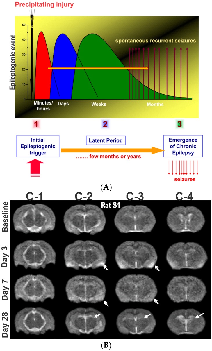
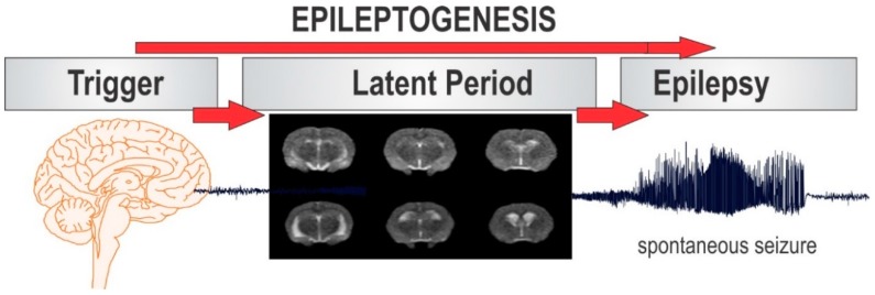
This article provides an overview of neuroimaging biomarkers in experimental epileptogenesis and refractory epilepsy. Neuroimaging represents a gold standard and clinically translatable technique to identify neuropathological changes in epileptogenesis and longitudinally monitor its progression after a precipitating injury. Neuroimaging studies, along with molecular studies from animal models, have greatly improved our understanding of the neuropathology of epilepsy, such as the hallmark hippocampus sclerosis. Animal models are effective for differentiating the different stages of epileptogenesis. Neuroimaging in experimental epilepsy provides unique information about anatomic, functional, and metabolic alterations linked to epileptogenesis. Recently, several in vivo biomarkers for epileptogenesis have been investigated for characterizing neuronal loss, inflammation, blood-brain barrier alterations, changes in neurotransmitter density, neurovascular coupling, cerebral blood flow and volume, network connectivity, and metabolic activity in the brain. Magnetic resonance imaging (MRI) is a sensitive method for detecting structural and functional changes in the brain, especially to identify region-specific neuronal damage patterns in epilepsy. Positron emission tomography (PET) and single-photon emission computerized tomography are helpful to elucidate key functional alterations, especially in areas of brain metabolism and molecular patterns, and can help monitor pathology of epileptic disorders. Multimodal procedures such as PET-MRI integrated systems are desired for refractory epilepsy. Validated biomarkers are warranted for early identification of people at risk for epilepsy and monitoring of the progression of medical interventions.
Keywords: MRI; PET; SPECT; biomarkers; epilepsy; epileptogenesis; imaging; seizures.
Conflict of interest statement
The authors declare no conflict of interest.
Figures
Similar articles
-
Neuroimaging in animal models of epilepsy.Neuroscience. 2017 Sep 1;358:277-299. doi: 10.1016/j.neuroscience.2017.06.062. Epub 2017 Jul 5. Neuroscience. 2017. PMID: 28688882 Review.
-
Altered intrinsic functional connectivity in the latent period of epileptogenesis in a temporal lobe epilepsy model.Exp Neurol. 2017 Oct;296:89-98. doi: 10.1016/j.expneurol.2017.07.007. Epub 2017 Jul 17. Exp Neurol. 2017. PMID: 28729114
-
Neuronuclear assessment of patients with epilepsy.Semin Nucl Med. 2008 Jul;38(4):227-39. doi: 10.1053/j.semnuclmed.2008.02.004. Semin Nucl Med. 2008. PMID: 18514079 Review.
-
Neuroimaging in epilepsy.Curr Opin Neurol. 2018 Aug;31(4):371-378. doi: 10.1097/WCO.0000000000000568. Curr Opin Neurol. 2018. PMID: 29782369 Review.
-
Positron Emission Tomography in basic epilepsy research: a view of the epileptic brain.Epilepsia. 2007;48 Suppl 4:56-64. doi: 10.1111/j.1528-1167.2007.01242.x. Epilepsia. 2007. PMID: 17767576
Cited by
-
Exploring with [18F]UCB-H the in vivo Variations in SV2A Expression through the Kainic Acid Rat Model of Temporal Lobe Epilepsy.Mol Imaging Biol. 2020 Oct;22(5):1197-1207. doi: 10.1007/s11307-020-01488-7. Mol Imaging Biol. 2020. PMID: 32206990 Free PMC article.
-
The Legacy of the TTASAAN Report - Premature Conclusions and Forgotten Promises About SPECT Neuroimaging: A Review of Policy and Practice Part II.Front Neurol. 2022 May 17;13:851609. doi: 10.3389/fneur.2022.851609. eCollection 2022. Front Neurol. 2022. PMID: 35655621 Free PMC article.
-
A longitudinal MRI and TSPO PET-based investigation of brain region-specific neuroprotection by diazepam versus midazolam following organophosphate-induced seizures.Neuropharmacology. 2024 Jun 15;251:109918. doi: 10.1016/j.neuropharm.2024.109918. Epub 2024 Mar 24. Neuropharmacology. 2024. PMID: 38527652 Free PMC article.
-
Post-Traumatic Epilepsy and Comorbidities: Advanced Models, Molecular Mechanisms, Biomarkers, and Novel Therapeutic Interventions.Pharmacol Rev. 2022 Apr;74(2):387-438. doi: 10.1124/pharmrev.121.000375. Pharmacol Rev. 2022. PMID: 35302046 Free PMC article. Review.
-
The Legacy of the TTASAAN Report-Premature Conclusions and Forgotten Promises: A Review of Policy and Practice Part I.Front Neurol. 2022 Mar 28;12:749579. doi: 10.3389/fneur.2021.749579. eCollection 2021. Front Neurol. 2022. PMID: 35450131 Free PMC article. Review.
References
Publication types
MeSH terms
Substances
Grants and funding
LinkOut - more resources
Full Text Sources
Medical
