

Osteoconductive resorption characteristics of a novel biocomposite suture anchor material in rotator cuff repair
- PMID: 30626411
- PMCID: PMC6325835
- DOI: 10.1186/s13018-018-1049-x
Osteoconductive resorption characteristics of a novel biocomposite suture anchor material in rotator cuff repair
Abstract
Background: Bioabsorbable suture anchors have been associated with bone-derived complications, such as osteolysis and cyst formation, after rotator cuff repair. The purpose of this study was to assess the osseous degradation process of the novel biocomposite suture anchor material polylactic-co-glycolic acid (PLGA)/beta-tricalcium phosphate (ß-TCP)/calcium sulfate (CS) after arthroscopic single-row rotator cuff repair. The focus of interest was the appearance of osteolysis and the rate of total resorption of the implants after 21 months.
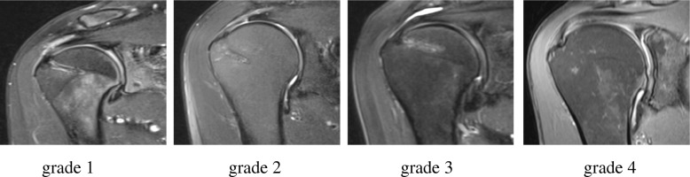
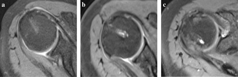
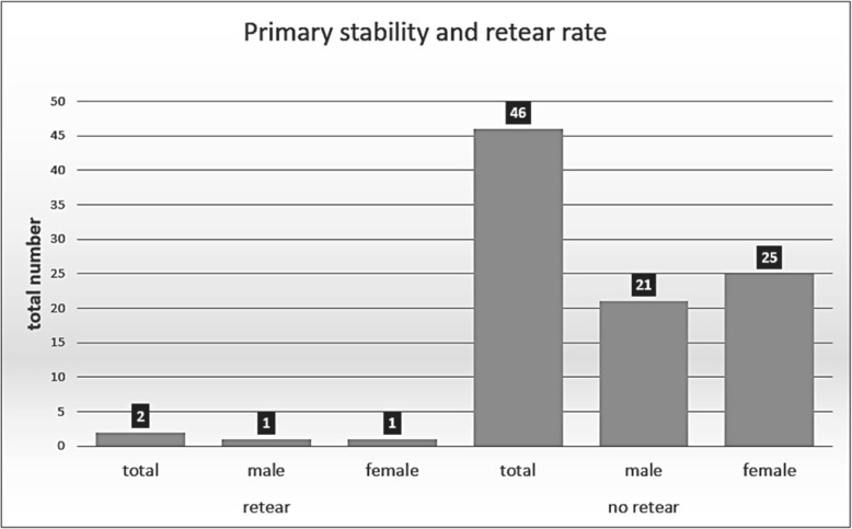
Methods: Forty-eight patients with 82 implanted suture anchors who had undergone arthroscopic rotator cuff repair between January 2015 and March 2016 at our institution were retrospectively evaluated by postoperative magnetic resonance imaging. The appearance of osteolysis was classified by measurement of the peri-implant fluid. The degree of resorption was measured by grading the persistent visibility of the anchor structures. The integrity of the rotator cuff tendon was analyzed to discover possible retear or anchor pull-out complications.
Results: After a follow-up of 21.2 (± 5.4) months, osteolysis was detected in only two anchors (2.4%), and none of these defects exceeded the diameter of the former suture anchor (5.5 mm). Fifty percent of the anchors were fully degraded and no longer visible. Furthermore, only two retears of the rotator cuff occurred, and no anchor pull-out complications were detected.
Conclusion: PGLA/β-TCP/CS is a fully resorbable and osteoconductive suture anchor material that seems to have superior resorption characteristics compared to those of other bioabsorbable suture anchor materials commonly used in arthroscopic rotator cuff repair.
Trial registration: The presented study was retrospectively registered by the commission for ethics at the Ärztekammer Nordrhein with the registration number 2016433 on January 17, 2017. All participating patients gave written consent for participation and the publication of their data.
Level of evidence: IV.
Keywords: Arthroscopic rotator cuff repair; Bioabsorbable; Osteolysis; PGLA; Suture anchor.
Conflict of interest statement
Ethics approval and consent to participate
The present study was retrospectively registered with the number 2016433 by the commission for ethics at the Ärztekammer Nordrhein on 17.01.2017. All participating patients gave written consent for participation.
Consent of publication
All participating patients gave written consent for participation and the publication of their data. All data were saved and acquired anonymously.
Competing interests
All authors declare that they have no competing interests.
Publisher’s Note
Springer Nature remains neutral with regard to jurisdictional claims in published maps and institutional affiliations.
Figures
Similar articles
-
Osteolysis is observed around both bioabsorbable and nonabsorbable anchors on serial magnetic resonance images of patients undergoing arthroscopic rotator cuff repair.Acta Orthop Traumatol Turc. 2019 Nov;53(6):414-419. doi: 10.1016/j.aott.2019.08.015. Epub 2019 Sep 25. Acta Orthop Traumatol Turc. 2019. PMID: 31563430 Free PMC article.
-
Biocomposite Suture Anchors Remain Visible Two Years After Rotator Cuff Repair.Clin Orthop Relat Res. 2019 Jun;477(6):1469-1478. doi: 10.1097/CORR.0000000000000665. Clin Orthop Relat Res. 2019. PMID: 30908350 Free PMC article.
-
The formation of perianchor fluid associated with various suture anchors used in rotator cuff repair: all-suture, polyetheretherketone, and biocomposite anchors.Bone Joint J. 2019 Dec;101-B(12):1506-1511. doi: 10.1302/0301-620X.101B12.BJJ-2019-0462.R2. Bone Joint J. 2019. PMID: 31786997 Clinical Trial.
-
Proximal humerus osteolysis after revision rotator cuff repair with bioabsorbable suture anchors.Am J Orthop (Belle Mead NJ). 2011 Mar;40(3):139-41. Am J Orthop (Belle Mead NJ). 2011. PMID: 21720602 Review.
-
Periimplant osteolysis does not affect the outcome of rotator cuff repair: a systematic review and meta-analysis.Knee Surg Sports Traumatol Arthrosc. 2021 Dec;29(12):3910-3920. doi: 10.1007/s00167-020-06328-3. Epub 2020 Oct 22. Knee Surg Sports Traumatol Arthrosc. 2021. PMID: 33090240
Cited by
-
Reliability of open architecture anchors in biocomposite material: medium term clinical and MRI evaluation. Our experience.Acta Biomed. 2020 May 30;91(4-S):189-195. doi: 10.23750/abm.v91i4-S.9709. Acta Biomed. 2020. PMID: 32555096 Free PMC article.
-
Biomechanical properties of a suture anchor system from human allogenic mineralized cortical bone matrix for rotator cuff repair.BMC Musculoskelet Disord. 2022 May 5;23(1):422. doi: 10.1186/s12891-022-05371-0. BMC Musculoskelet Disord. 2022. PMID: 35513813 Free PMC article.
-
Protocol for a Retrospective Comparative Study to Determine the Effect of Two Different Biocomposite Suture Anchors on the Occurrence of Bony Ingrowth and Implant Reabsorption Following Arthroscopic Rotator Cuff Repair.Int J Surg Protoc. 2021 Jul 29;25(1):147-153. doi: 10.29337/ijsp.140. eCollection 2021. Int J Surg Protoc. 2021. PMID: 34395961 Free PMC article.
-
Comparison of All-Suture Anchors and Metal Anchors in Arthroscopic Rotator Cuff Repair: Short-Term Clinical Outcomes and Anchor Pullout Risk.J Clin Med. 2025 Apr 11;14(8):2619. doi: 10.3390/jcm14082619. J Clin Med. 2025. PMID: 40283449 Free PMC article.
-
Absorbable implants in sport medicine and arthroscopic surgery: A narrative review of recent development.Bioact Mater. 2023 Aug 17;31:272-283. doi: 10.1016/j.bioactmat.2023.08.015. eCollection 2024 Jan. Bioact Mater. 2023. PMID: 37637087 Free PMC article. Review.
References
-
- Oliva F, Piccirilli E, Bossa M, Via AG, Colombo A, Chillemi C, Gasparre G, Pellicciari L, Franceschetti E, Rugiero C, Scialdoni A, Vittadini F, Brancaccio P, Creta D, Buono AD, Garofalo R, Franceschi F, Frizziero A, Mahmoud A, Merolla G, Nicoletti S, Spoliti M, Osti L, Padulo J, Portinaro N, Tajana G, Castagna A, Foti C, Masiero S, Porcellini G, Tarantino U, Maffulli NISMLT. Rotator cuff tears guidelines. Muscle Ligaments Tendons J. 2016;5(4):227–263. - PMC - PubMed
MeSH terms
Substances
LinkOut - more resources
Full Text Sources
Medical
Research Materials
